7P5R

Racemic protein crystal structure of lacticin Q from Lactococcus lactis


Experimental Data Snapshot

  • Method: X-RAY DIFFRACTION
  • Resolution: 0.96 Å
  • R-Value Free: 0.205 
  • R-Value Work: 0.182 

wwPDB Validation   3D Report Full Report


This is version 1.2 of the entry. See complete history


Literature

Roles of inter- and intramolecular tryptophan interactions in membrane-active proteins revealed by racemic protein crystallography.

Lander, A.J.Mercado, L.D.Li, X.Taily, I.M.Findlay, B.L.Jin, Y.Luk, L.Y.P.

(2023) Commun Chem 6: 154-154

  • DOI: https://doi.org/10.1038/s42004-023-00953-y
  • Primary Citation of Related Structures:  
    7P5R, 8AVR, 8AVS, 8AVT, 8AVU

  • PubMed Abstract: 

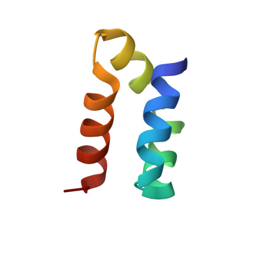
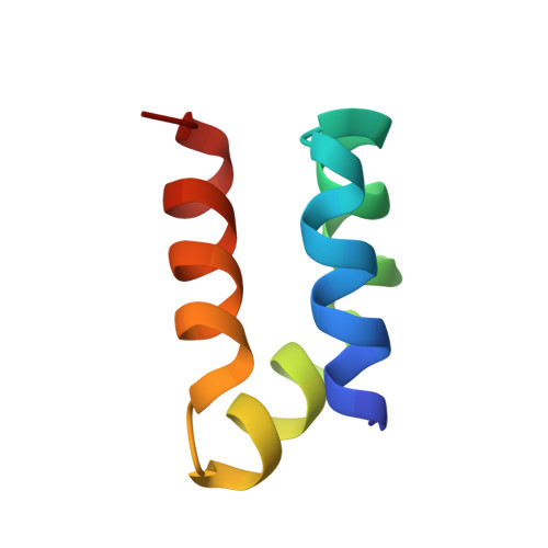
    Tryptophan is frequently found on the surface of membrane-associated proteins that interact with the lipid membrane. However, because of their multifaceted interactions, it is difficult to pinpoint the structure-activity relationship of each tryptophan residue. Here, we describe the use of racemic protein crystallography to probe dedicated tryptophan interactions of a model tryptophan-rich bacteriocin aureocin A53 (AucA) by inclusion and/or exclusion of potential ligands. In the presence of tetrahedral anions that are isosteric to the head group of phospholipids, distinct tryptophan H-bond networks were revealed. H-bond donation by W40 was critical for antibacterial activity, as its substitution by 1-methyltryptophan resulted in substantial loss of activity against bacterial clinical isolates. Meanwhile, exclusion of tetrahedral ions revealed that W3 partakes in formation of a dimeric interface, thus suggesting that AucA is dimeric in solution and dissociated to interact with the phosphate head group in the presence of the lipid membrane. Based on these findings, we could predict the tryptophan residue responsible for activity as well as the oligomeric state of a distant homologue lacticin Q (48%).


  • Organizational Affiliation

    School of Chemistry, Cardiff University, Main Building, Park Place, Cardiff, CF10 3AT, UK.


Macromolecules
Find similar proteins by:  (by identity cutoff)  |  3D Structure
Entity ID: 1
MoleculeChains Sequence LengthOrganismDetailsImage
Lacticin QA [auth AAA],
B [auth BBB]
53Lactococcus lactisMutation(s): 0 
UniProt
Find proteins for A4UVR2 (Lactococcus lactis)
Explore A4UVR2 
Go to UniProtKB:  A4UVR2
Entity Groups  
Sequence Clusters30% Identity50% Identity70% Identity90% Identity95% Identity100% Identity
UniProt GroupA4UVR2
Sequence Annotations
Expand
  • Reference Sequence
Find similar proteins by:  (by identity cutoff)  |  3D Structure
Entity ID: 2
MoleculeChains Sequence LengthOrganismDetailsImage
D-Lacticin QC [auth DDD],
D [auth CCC]
53Lactococcus lactisMutation(s): 0 
Entity Groups  
Sequence Clusters30% Identity50% Identity70% Identity90% Identity95% Identity100% Identity
Sequence Annotations
Expand
  • Reference Sequence
Experimental Data & Validation

Experimental Data

  • Method: X-RAY DIFFRACTION
  • Resolution: 0.96 Å
  • R-Value Free: 0.205 
  • R-Value Work: 0.182 
  • Space Group: P 1
Unit Cell:
Length ( Å )Angle ( ˚ )
a = 24.21α = 95.03
b = 27.83β = 90.43
c = 70.45γ = 115.41
Software Package:
Software NamePurpose
REFMACrefinement
Aimlessdata scaling
Fragonphasing
Cootmodel building
SHELXEmodel building
XDSdata reduction
xia2data reduction

Structure Validation

View Full Validation Report


Entry History & Funding Information

Deposition Data


Funding OrganizationLocationGrant Number
Engineering and Physical Sciences Research CouncilUnited KingdomEP/L027240/1
Biotechnology and Biological Sciences Research Council (BBSRC)United KingdomBB/T015799/1
Leverhulme TrustUnited KingdomRPG-2017-195
Royal SocietyUnited KingdomRG170187
Engineering and Physical Sciences Research CouncilUnited Kingdom2107414

Revision History  (Full details and data files)

  • Version 1.0: 2022-07-27
    Type: Initial release
  • Version 1.1: 2023-07-26
    Changes: Database references, Structure summary
  • Version 1.2: 2024-05-01
    Changes: Data collection, Refinement description