7P53 | pdb_00007p53

Crystal Structure of Human gamma-D-crystallin mutant C110M at 1.57 Angstroms resolution


Experimental Data Snapshot

  • Method: X-RAY DIFFRACTION
  • Resolution: 1.57 Å
  • R-Value Free: 
    0.274 (Depositor), 0.271 (DCC) 
  • R-Value Work: 
    0.215 (Depositor), 0.221 (DCC) 
  • R-Value Observed: 
    0.217 (Depositor) 

Starting Model: experimental
View more details

wwPDB Validation 3D Report Full Report

Validation slider image for 7P53

This is version 1.1 of the entry. See complete history

Literature

Surface Exposed Free Cysteine Suppresses Crystallization of Human gamma D-Crystallin.

Strofaldi, A.Khan, A.R.McManus, J.J.

(2021) J Mol Biology 433: 167252-167252

  • DOI: https://doi.org/10.1016/j.jmb.2021.167252
  • Primary Citation Related Structures: 
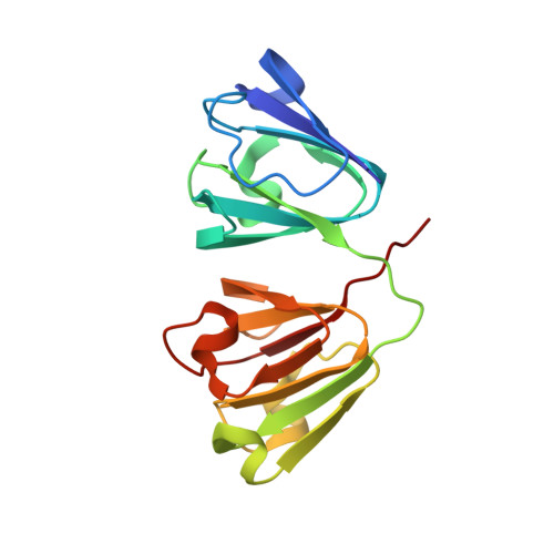
    7P53

  • PubMed Abstract: 

    Human γD-crystallin (HGD) has remarkable stability against condensation in the human lens, sometimes over a whole lifetime. The native protein has a surface exposed free cysteine that forms dimers (Benedek, 1997; Ramkumar et al., 1864) 1,2 without specific biological function and leads to further protein association and/or aggregation, which creates a paradox for understanding its stability. Previous work has demonstrated that chemical modification of the protein at the free cysteine (C110), increases the temperature at which liquid-liquid phase separation occurs (LLPS), lowers protein solubility and suggests an important role for this amino acid in maintaining its long-term resistance to condensation. Here we demonstrate that mutation of the cysteine does not alter the structure or solubility (liquidus) line for the protein, but dramatically increases the protein crystal nucleation rate following LLPS, suggesting that the free cysteine has a vital role in suppressing crystallization in the human lens.


  • Organizational Affiliation
    • Department of Chemistry, Maynooth University, Maynooth, Co. Kildare, Ireland; H. H Wills Physics Laboratory, University of Bristol, Bristol BS8 1TL, United Kingdom.

Macromolecule Content 

  • Total Structure Weight: 20.66 kDa 
  • Atom Count: 1,575 
  • Modeled Residue Count: 173 
  • Deposited Residue Count: 173 
  • Unique protein chains: 1

Macromolecules

Find similar proteins by:|  3D Structure
Entity ID: 1
MoleculeChains  Sequence LengthOrganismDetailsImage
Gamma-crystallin DA [auth X]173Homo sapiensMutation(s): 1 
Gene Names: CRYGDCRYG4
UniProt & NIH Common Fund Data Resources
Find proteins for P07320 (Homo sapiens)
Explore P07320 
Go to UniProtKB:  P07320
PHAROS:  P07320
GTEx:  ENSG00000118231 
Entity Groups
Sequence Clusters30% Identity50% Identity70% Identity90% Identity95% Identity100% Identity
UniProt GroupP07320
Sequence Annotations
Expand
Reference Sequence

Experimental Data & Validation

Experimental Data

  • Method: X-RAY DIFFRACTION
  • Resolution: 1.57 Å
  • R-Value Free:  0.274 (Depositor), 0.271 (DCC) 
  • R-Value Work:  0.215 (Depositor), 0.221 (DCC) 
  • R-Value Observed: 0.217 (Depositor) 
Space Group: P 21 21 21
Unit Cell:
Length ( Å )Angle ( ˚ )
a = 33.699α = 90
b = 52.844β = 90
c = 89.554γ = 90
Software Package:
Software NamePurpose
PHENIXrefinement
PDB_EXTRACTdata extraction
XDSdata reduction
Aimlessdata scaling
PHENIXphasing

Structure Validation

View Full Validation Report


Entry History 

& Funding Information

Deposition Data


Funding OrganizationLocationGrant Number
Other governmentIrelandBU1300037

Revision History  (Full details and data files)

  • Version 1.0: 2021-10-13
    Type: Initial release
  • Version 1.1: 2024-01-31
    Changes: Data collection, Refinement description