7P4B

HLA-E*01:03 in complex with IL9


Experimental Data Snapshot

  • Method: X-RAY DIFFRACTION
  • Resolution: 1.72 Å
  • R-Value Free: 0.237 
  • R-Value Work: 0.201 
  • R-Value Observed: 0.203 

wwPDB Validation   3D Report Full Report


This is version 1.2 of the entry. See complete history


Literature

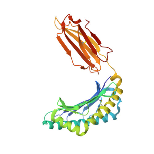
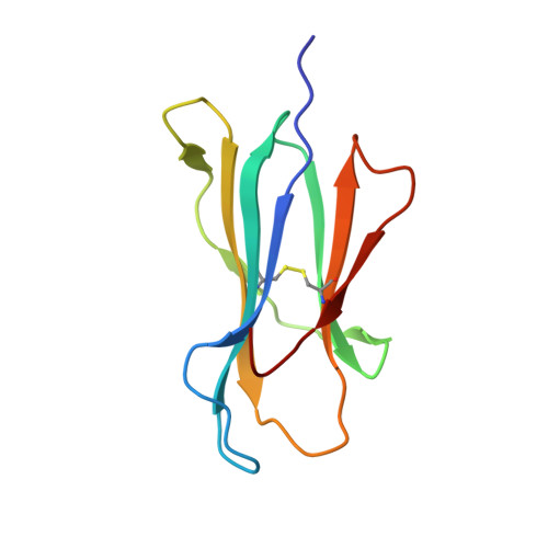
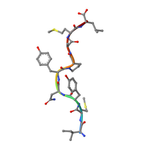
Primary and secondary functions of HLA-E are determined by stability and conformation of the peptide-bound complexes.

Walters, L.C.Rozbesky, D.Harlos, K.Quastel, M.Sun, H.Springer, S.Rambo, R.P.Mohammed, F.Jones, E.Y.McMichael, A.J.Gillespie, G.M.

(2022) Cell Rep 39: 110959-110959

  • DOI: https://doi.org/10.1016/j.celrep.2022.110959
  • Primary Citation of Related Structures:  
    7P49, 7P4B

  • PubMed Abstract: 

    MHC-E regulates NK cells by displaying MHC class Ia signal peptides (VL9) to NKG2A:CD94 receptors. MHC-E can also present sequence-diverse, lower-affinity, pathogen-derived peptides to T cell receptors (TCRs) on CD8 + T cells. To understand these affinity differences, human MHC-E (HLA-E)-VL9 versus pathogen-derived peptide structures are compared. Small-angle X-ray scatter (SAXS) measures biophysical parameters in solution, allowing comparison with crystal structures. For HLA-E-VL9, there is concordance between SAXS and crystal parameters. In contrast, HLA-E-bound pathogen-derived peptides produce larger SAXS dimensions that reduce to their crystallographic dimensions only when excess peptide is supplied. Further crystallographic analysis demonstrates three amino acids, exclusive to MHC-E, that not only position VL9 close to the α2 helix, but also allow non-VL9 peptide binding with re-configuration of a key TCR-interacting α2 region. Thus, non-VL9-bound peptides introduce an alternative peptide-binding motif and surface recognition landscape, providing a likely basis for VL9- and non-VL9-HLA-E immune discrimination.


  • Organizational Affiliation

    Nuffield Department of Medicine Research Building, Nuffield Department of Medicine, University of Oxford, Roosevelt Drive, Oxford OX3 7FZ, UK.


Macromolecules
Find similar proteins by:  (by identity cutoff)  |  3D Structure
Entity ID: 1
MoleculeChains Sequence LengthOrganismDetailsImage
HLA class I histocompatibility antigen, alpha chain E
A, C, E, G
277Homo sapiensMutation(s): 0 
Gene Names: HLA-EHLA-6.2HLAE
UniProt & NIH Common Fund Data Resources
Find proteins for P13747 (Homo sapiens)
Explore P13747 
Go to UniProtKB:  P13747
PHAROS:  P13747
GTEx:  ENSG00000204592 
Entity Groups  
Sequence Clusters30% Identity50% Identity70% Identity90% Identity95% Identity100% Identity
UniProt GroupP13747
Sequence Annotations
Expand
  • Reference Sequence
Find similar proteins by:  (by identity cutoff)  |  3D Structure
Entity ID: 2
MoleculeChains Sequence LengthOrganismDetailsImage
Beta-2-microglobulin
B, D, F, H
100Homo sapiensMutation(s): 0 
Gene Names: B2MCDABP0092HDCMA22P
UniProt & NIH Common Fund Data Resources
Find proteins for P61769 (Homo sapiens)
Explore P61769 
Go to UniProtKB:  P61769
PHAROS:  P61769
GTEx:  ENSG00000166710 
Entity Groups  
Sequence Clusters30% Identity50% Identity70% Identity90% Identity95% Identity100% Identity
UniProt GroupP61769
Sequence Annotations
Expand
  • Reference Sequence

Find similar proteins by:  Sequence   |   3D Structure  

Entity ID: 3
MoleculeChains Sequence LengthOrganismDetailsImage
ESAT-6-like protein EsxHI [auth P],
J [auth Q],
K [auth R],
L [auth Z]
9Mycobacterium tuberculosis H37RvMutation(s): 0 
Gene Names: esxHcfp7Rv0288MTV035.16
UniProt
Find proteins for P9WNK3 (Mycobacterium tuberculosis (strain ATCC 25618 / H37Rv))
Explore P9WNK3 
Go to UniProtKB:  P9WNK3
Entity Groups  
UniProt GroupP9WNK3
Sequence Annotations
Expand
  • Reference Sequence
Small Molecules
Ligands 4 Unique
IDChains Name / Formula / InChI Key2D Diagram3D Interactions
SO4
Query on SO4

Download Ideal Coordinates CCD File 
AA [auth D]
CA [auth F]
DA [auth F]
HA [auth G]
IA [auth G]
AA [auth D],
CA [auth F],
DA [auth F],
HA [auth G],
IA [auth G],
JA [auth G],
LA [auth H],
MA [auth H],
N [auth A],
NA [auth H],
O [auth A],
OA [auth H],
P [auth A],
S [auth C],
T [auth C],
U [auth C]
SULFATE ION
O4 S
QAOWNCQODCNURD-UHFFFAOYSA-L
PO4
Query on PO4

Download Ideal Coordinates CCD File 
EA [auth F]PHOSPHATE ION
O4 P
NBIIXXVUZAFLBC-UHFFFAOYSA-K
GOL
Query on GOL

Download Ideal Coordinates CCD File 
FA [auth G]
KA [auth H]
M [auth A]
Q [auth C]
R [auth C]
FA [auth G],
KA [auth H],
M [auth A],
Q [auth C],
R [auth C],
V [auth C],
W [auth D],
X [auth D],
Y [auth D],
Z [auth D]
GLYCEROL
C3 H8 O3
PEDCQBHIVMGVHV-UHFFFAOYSA-N
ZN
Query on ZN

Download Ideal Coordinates CCD File 
BA [auth E],
GA [auth G]
ZINC ION
Zn
PTFCDOFLOPIGGS-UHFFFAOYSA-N
Experimental Data & Validation

Experimental Data

  • Method: X-RAY DIFFRACTION
  • Resolution: 1.72 Å
  • R-Value Free: 0.237 
  • R-Value Work: 0.201 
  • R-Value Observed: 0.203 
  • Space Group: C 1 2 1
Unit Cell:
Length ( Å )Angle ( ˚ )
a = 244.562α = 90
b = 48.529β = 116.948
c = 153.161γ = 90
Software Package:
Software NamePurpose
PHENIXrefinement
PHENIXrefinement
xia2data reduction
xia2data scaling
MOLREPphasing

Structure Validation

View Full Validation Report


Entry History & Funding Information

Deposition Data


Funding OrganizationLocationGrant Number
Bill & Melinda Gates Foundation--

Revision History  (Full details and data files)

  • Version 1.0: 2022-07-27
    Type: Initial release
  • Version 1.1: 2022-08-03
    Changes: Database references
  • Version 1.2: 2024-01-31
    Changes: Data collection, Refinement description