7P34

Cryo-EM structure of the proton-dependent antibacterial peptide transporter SbmA-FabS11-1 in nanodiscs


Experimental Data Snapshot

  • Method: ELECTRON MICROSCOPY
  • Resolution: 3.59 Å
  • Aggregation State: PARTICLE 
  • Reconstruction Method: SINGLE PARTICLE 

wwPDB Validation   3D Report Full Report


This is version 1.1 of the entry. See complete history


Literature

Molecular mechanism of SbmA, a promiscuous transporter exploited by antimicrobial peptides.

Ghilarov, D.Inaba-Inoue, S.Stepien, P.Qu, F.Michalczyk, E.Pakosz, Z.Nomura, N.Ogasawara, S.Walker, G.C.Rebuffat, S.Iwata, S.Heddle, J.G.Beis, K.

(2021) Sci Adv 7: eabj5363-eabj5363

  • DOI: https://doi.org/10.1126/sciadv.abj5363
  • Primary Citation of Related Structures:  
    7P34

  • PubMed Abstract: 

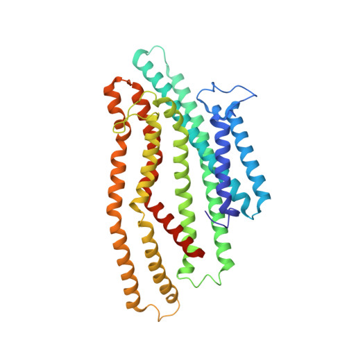
    Antibiotic metabolites and antimicrobial peptides mediate competition between bacterial species. Many of them hijack inner and outer membrane proteins to enter cells. Sensitivity of enteric bacteria to multiple peptide antibiotics is controlled by the single inner membrane protein SbmA. To establish the molecular mechanism of peptide transport by SbmA and related BacA, we determined their cryo–electron microscopy structures at 3.2 and 6 Å local resolution, respectively. The structures show a previously unknown fold, defining a new class of secondary transporters named SbmA-like peptide transporters. The core domain includes conserved glutamates, which provide a pathway for proton translocation, powering transport. The structures show an outward-open conformation with a large cavity that can accommodate diverse substrates. We propose a molecular mechanism for antibacterial peptide uptake paving the way for creation of narrow-targeted therapeutics.


  • Organizational Affiliation

    Malopolska Centre of Biotechnology, Jagiellonian University, Krakow, Poland.


Macromolecules
Find similar proteins by:  (by identity cutoff)  |  3D Structure
Entity ID: 1
MoleculeChains Sequence LengthOrganismDetailsImage
Peptide antibiotic transporter SbmA
A, B
406Escherichia coliMutation(s): 0 
Membrane Entity: Yes 
UniProt
Find proteins for P0AFY6 (Escherichia coli (strain K12))
Explore P0AFY6 
Go to UniProtKB:  P0AFY6
Entity Groups  
Sequence Clusters30% Identity50% Identity70% Identity90% Identity95% Identity100% Identity
UniProt GroupP0AFY6
Sequence Annotations
Expand
  • Reference Sequence
Small Molecules
Ligands 1 Unique
IDChains Name / Formula / InChI Key2D Diagram3D Interactions
PGW
Query on PGW

Download Ideal Coordinates CCD File 
C [auth A],
D [auth B]
(1R)-2-{[(S)-{[(2S)-2,3-dihydroxypropyl]oxy}(hydroxy)phosphoryl]oxy}-1-[(hexadecanoyloxy)methyl]ethyl (9Z)-octadec-9-enoate
C40 H77 O10 P
PAZGBAOHGQRCBP-HGWHEPCSSA-N
Experimental Data & Validation

Experimental Data

  • Method: ELECTRON MICROSCOPY
  • Resolution: 3.59 Å
  • Aggregation State: PARTICLE 
  • Reconstruction Method: SINGLE PARTICLE 
EM Software:
TaskSoftware PackageVersion
RECONSTRUCTIONcryoSPARC3.2.0
MODEL REFINEMENTPHENIX

Structure Validation

View Full Validation Report


Entry History & Funding Information

Deposition Data


Funding OrganizationLocationGrant Number
Biotechnology and Biological Sciences Research Council (BBSRC)United KingdomBB/H01778X/1
Japan Society for the Promotion of Science (JSPS)Japan--
Foundation for Polish SciencePolandTEAM/2016-3/23
Polish National Science CentrePoland2016/21/B/CC1/00274 (OPUS 11)
Polish National Science CentrePoland2019/35/D/NZ1/01770 (SONATA 15)

Revision History  (Full details and data files)

  • Version 1.0: 2021-09-15
    Type: Initial release
  • Version 1.1: 2021-09-29
    Changes: Data collection, Database references, Structure summary