7P2C | pdb_00007p2c

F(M197)H mutant structure of Photosynthetic Reaction Center From Rhodobacter Sphaeroides strain RV by fixed-target serial synchrotron crystallography (room temperature, 26keV)


Experimental Data Snapshot

  • Method: X-RAY DIFFRACTION
  • Resolution: 2.04 Å
  • R-Value Free: 
    0.229 (Depositor), 0.241 (DCC) 
  • R-Value Work: 
    0.201 (Depositor), 0.211 (DCC) 
  • R-Value Observed: 
    0.202 (Depositor) 

Starting Model: experimental
View more details

wwPDB Validation 3D Report Full Report

Validation slider image for 7P2C

Ligand Structure Quality Assessment 


This is version 1.1 of the entry. See complete history

Literature

X-ray structure of the Rhodobacter sphaeroides reaction center with an M197 Phe→His substitution clarifies the properties of the mutant complex.

Selikhanov, G.Fufina, T.Guenther, S.Meents, A.Gabdulkhakov, A.Vasilieva, L.

(2022) IUCrJ 9: 261-271

  • DOI: https://doi.org/10.1107/S2052252521013178
  • Primary Citation Related Structures: 
    7OD5, 7P2C

  • PubMed Abstract: 

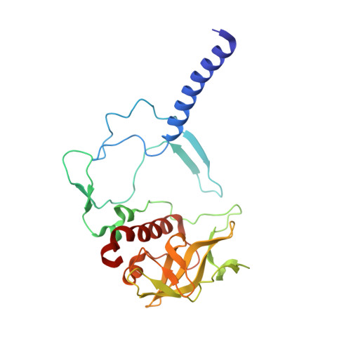
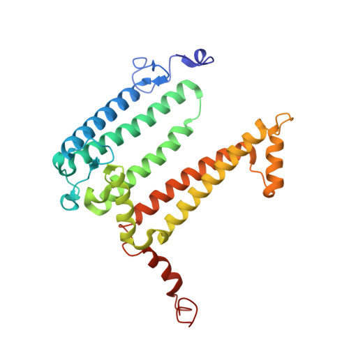
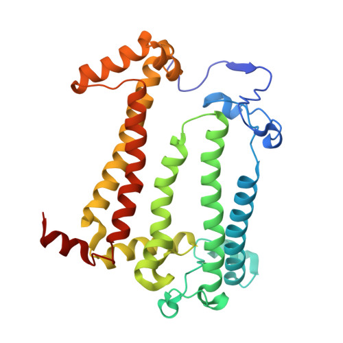
    The first steps of the global process of photosynthesis take place in specialized membrane pigment-protein complexes called photosynthetic reaction centers (RCs). The RC of the photosynthetic purple bacterium Rhodobacter sphaeroides , a relatively simple analog of the more complexly organized photosystem II in plants, algae and cyanobacteria, serves as a convenient model for studying pigment-protein interactions that affect photochemical processes. In bacterial RCs the bacteriochlorophyll (BChl) dimer P serves as the primary electron donor, and its redox potential is a critical factor in the efficient functioning of the RC. It has previously been shown that the replacement of Phe M197 by His strongly affects the oxidation potential of P ( E m P/P + ), increasing its value by 125 mV, as well as increasing the thermal stability of RC and its stability in response to external pressure. The crystal structures of F(M197)H RC at high resolution obtained using various techniques presented in this report clarify the optical and electrochemical properties of the primary electron donor and the increased resistance of the mutant complex to denaturation. The electron-density maps are consistent with the donation of a hydrogen bond from the imidazole group of His M197 to the C2-acetyl carbonyl group of BChl P B . The formation of this hydrogen bond leads to a considerable out-of-plane rotation of the acetyl carbonyl group and results in a 1.2 Å shift of the O atom of this group relative to the wild-type structure. Besides, the distance between BChl P A and P B in the area of pyrrole ring I was found to be increased by up to 0.17 Å. These structural changes are discussed in association with the spectral properties of BChl dimer P. The electron-density maps strongly suggest that the imidazole group of His M197 accepts another hydrogen bond from the nearest water molecule, which in turn appears to form two more hydrogen bonds to Asn M195 and Asp L155. As a result of the F(M197)H mutation, BChl P B finds itself connected to the extensive hydrogen-bonding network that pre-existed in wild-type RC. Dissimilarities in the two hydrogen-bonding networks near the M197 and L168 sites may account for the different changes of the E m P/P + in F(M197)H and H(L168)F RCs. The involvement of His M197 in the hydrogen-bonding network also appears to be related to stabilization of the F(M197)H RC structure. Analysis of the experimental data presented here and of the data available in the literature points to the fact that the hydrogen-bonding networks in the vicinity of BChl dimer P may play an important role in fine-tuning the redox properties of the primary electron donor.


  • Organizational Affiliation
    • Group of Structural Studies of Macromolecular Complexes, Institute of Protein Research, Russian Academy of Sciences, Institutskaya 4, Pushchino 142290, Moscow Region, Russian Federation.

Macromolecule Content 

  • Total Structure Weight: 101.07 kDa 
  • Atom Count: 7,591 
  • Modeled Residue Count: 822 
  • Deposited Residue Count: 826 
  • Unique protein chains: 3

Macromolecules

Find similar proteins by:|  3D Structure
Entity ID: 1
MoleculeChains  Sequence LengthOrganismDetailsImage
Reaction center protein H chainA [auth H]242Cereibacter sphaeroides 2.4.1Mutation(s): 0 
Gene Names: puhARHOS4_18960RSP_0291
Membrane Entity: Yes 
UniProt
Find proteins for Q3J170 (Cereibacter sphaeroides (strain ATCC 17023 / DSM 158 / JCM 6121 / CCUG 31486 / LMG 2827 / NBRC 12203 / NCIMB 8253 / ATH 2.4.1.))
Explore Q3J170 
Go to UniProtKB:  Q3J170
Entity Groups
Sequence Clusters30% Identity50% Identity70% Identity90% Identity95% Identity100% Identity
UniProt GroupQ3J170
Sequence Annotations
Expand
Reference Sequence
Find similar proteins by:|  3D Structure
Entity ID: 2
MoleculeChains  Sequence LengthOrganismDetailsImage
Reaction center protein L chainB [auth L]281Cereibacter sphaeroides 2.4.1Mutation(s): 1 
Gene Names: pufLRHOS4_18610RSP_0257
Membrane Entity: Yes 
UniProt
Find proteins for Q3J1A5 (Cereibacter sphaeroides (strain ATCC 17023 / DSM 158 / JCM 6121 / CCUG 31486 / LMG 2827 / NBRC 12203 / NCIMB 8253 / ATH 2.4.1.))
Explore Q3J1A5 
Go to UniProtKB:  Q3J1A5
Entity Groups
Sequence Clusters30% Identity50% Identity70% Identity90% Identity95% Identity100% Identity
UniProt GroupQ3J1A5
Sequence Annotations
Expand
Reference Sequence
Find similar proteins by:|  3D Structure
Entity ID: 3
MoleculeChains  Sequence LengthOrganismDetailsImage
Reaction center protein M chainC [auth M]303Cereibacter sphaeroides 2.4.1Mutation(s): 2 
Gene Names: pufMRHOS4_18600RSP_0256
Membrane Entity: Yes 
UniProt
Find proteins for Q3J1A6 (Cereibacter sphaeroides (strain ATCC 17023 / DSM 158 / JCM 6121 / CCUG 31486 / LMG 2827 / NBRC 12203 / NCIMB 8253 / ATH 2.4.1.))
Explore Q3J1A6 
Go to UniProtKB:  Q3J1A6
Entity Groups
Sequence Clusters30% Identity50% Identity70% Identity90% Identity95% Identity100% Identity
UniProt GroupQ3J1A6
Sequence Annotations
Expand
Reference Sequence

Small Molecules

Ligands 11 Unique
IDChains Name / Formula / InChI Key2D Diagram3D Interactions
BCL
(Subject of Investigation/LOI)

Query on BCL


Download:Ideal Coordinates CCD File
H [auth L],
I [auth L],
N [auth M],
O [auth M]
BACTERIOCHLOROPHYLL A
C55 H74 Mg N4 O6
DSJXIQQMORJERS-AGGZHOMASA-M
BPH
(Subject of Investigation/LOI)

Query on BPH


Download:Ideal Coordinates CCD File
J [auth L],
P [auth M]
BACTERIOPHEOPHYTIN A
C55 H76 N4 O6
KWOZSBGNAHVCKG-SZQBJALDSA-N
U10

Query on U10


Download:Ideal Coordinates CCD File
L [auth M]UBIQUINONE-10
C59 H90 O4
ACTIUHUUMQJHFO-UPTCCGCDSA-N
SPN

Query on SPN


Download:Ideal Coordinates CCD File
R [auth M]SPEROIDENONE
C41 H70 O2
GWQAMGYOEYXWJF-YCDPMLDASA-N
NKP

Query on NKP


Download:Ideal Coordinates CCD File
K [auth M],
M
(2R)-2-hydroxy-3-(phosphonooxy)propyl (9E)-octadec-9-enoate
C21 H41 O7 P
WRGQSWVCFNIUNZ-SQUSKLHYSA-N
OLC

Query on OLC


Download:Ideal Coordinates CCD File
E [auth L](2R)-2,3-dihydroxypropyl (9Z)-octadec-9-enoate
C21 H40 O4
RZRNAYUHWVFMIP-GDCKJWNLSA-N
LDA

Query on LDA


Download:Ideal Coordinates CCD File
D [auth H],
S [auth M]
LAURYL DIMETHYLAMINE-N-OXIDE
C14 H31 N O
SYELZBGXAIXKHU-UHFFFAOYSA-N
MYS

Query on MYS


Download:Ideal Coordinates CCD File
F [auth L],
G [auth L],
T [auth M]
PENTADECANE
C15 H32
YCOZIPAWZNQLMR-UHFFFAOYSA-N
PO4

Query on PO4


Download:Ideal Coordinates CCD File
V [auth M]PHOSPHATE ION
O4 P
NBIIXXVUZAFLBC-UHFFFAOYSA-K
EDO

Query on EDO


Download:Ideal Coordinates CCD File
U [auth M]1,2-ETHANEDIOL
C2 H6 O2
LYCAIKOWRPUZTN-UHFFFAOYSA-N
FE

Query on FE


Download:Ideal Coordinates CCD File
Q [auth M]FE (III) ION
Fe
VTLYFUHAOXGGBS-UHFFFAOYSA-N

Experimental Data & Validation

Experimental Data

  • Method: X-RAY DIFFRACTION
  • Resolution: 2.04 Å
  • R-Value Free:  0.229 (Depositor), 0.241 (DCC) 
  • R-Value Work:  0.201 (Depositor), 0.211 (DCC) 
  • R-Value Observed: 0.202 (Depositor) 
Space Group: P 42 21 2
Unit Cell:
Length ( Å )Angle ( ˚ )
a = 102.5α = 90
b = 102.5β = 90
c = 237.4γ = 90
Software Package:
Software NamePurpose
REFMACrefinement
PHENIXrefinement
XDSdata reduction
XSCALEdata scaling
PHASERphasing

Structure Validation

View Full Validation Report


Ligand Structure Quality Assessment 


Entry History 

& Funding Information

Deposition Data


Funding OrganizationLocationGrant Number
Russian Foundation for Basic ResearchRussian Federation18-02-40008_mega

Revision History  (Full details and data files)

  • Version 1.0: 2022-04-27
    Type: Initial release
  • Version 1.1: 2024-01-31
    Changes: Data collection, Refinement description