7P18

Crystal structure of 3-ketosteroid delta1-dehydrogenase from Sterolibacterium denitrificans in complex with 1,4-androstadiene-3,17-dione


Experimental Data Snapshot

  • Method: X-RAY DIFFRACTION
  • Resolution: 1.84 Å
  • R-Value Free: 0.214 
  • R-Value Work: 0.185 
  • R-Value Observed: 0.185 

wwPDB Validation   3D Report Full Report


Ligand Structure Quality Assessment 


This is version 1.4 of the entry. See complete history


Literature

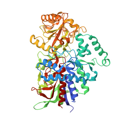
Structure, Mutagenesis, and QM:MM Modeling of 3-Ketosteroid Delta 1 -Dehydrogenase from Sterolibacterium denitrificans ─The Role of a New Putative Membrane-Associated Domain and Proton-Relay System in Catalysis.

Wojcik, P.Glanowski, M.Mrugala, B.Procner, M.Zastawny, O.Flejszar, M.Kurpiewska, K.Niedzialkowska, E.Minor, W.Oszajca, M.Bojarski, A.J.Wojtkiewicz, A.M.Szaleniec, M.

(2023) Biochemistry 62: 808-823

  • DOI: https://doi.org/10.1021/acs.biochem.2c00576
  • Primary Citation of Related Structures:  
    7P18

  • PubMed Abstract: 

    3-Ketosteroid Δ 1 -dehydrogenases (KstD) are important microbial flavin enzymes that initiate the metabolism of steroid ring A and find application in the synthesis of steroid drugs. We present a structure of the KstD from Sterolibacterium denitrificans (AcmB), which contains a previously uncharacterized putative membrane-associated domain and extended proton-relay system. The experimental and theoretical studies show that the steroid Δ 1 -dehydrogenation proceeds according to the Ping-Pong bi-bi kinetics and a two-step base-assisted elimination (E2cB) mechanism. The mechanism is validated by evaluating the experimental and theoretical kinetic isotope effect for deuterium-substituted substrates. The role of the active-site residues is quantitatively assessed by point mutations, experimental activity assays, and QM/MM MD modeling of the reductive half-reaction (RHR). The pre-steady-state kinetics also reveals that the low pH (6.5) optimum of AcmB is dictated by the oxidative half-reaction (OHR), while the RHR exhibits a slight optimum at the pH usual for the KstD family of 8.5. The modeling confirms the origin of the enantioselectivity of C2-H activation and substrate specificity for Δ 4 -3-ketosteroids. Finally, the cholest-4-en-3-one turns out to be the best substrate of AcmB in terms of Δ G of binding and predicted rate of dehydrogenation.


  • Organizational Affiliation

    Jerzy Haber Institute of Catalysis and Surface Chemistry, Polish Academy of Sciences, Niezapominajek 8, 30-239Kraków, Poland.


Macromolecules
Find similar proteins by:  (by identity cutoff)  |  3D Structure
Entity ID: 1
MoleculeChains Sequence LengthOrganismDetailsImage
3-oxosteroid 1-dehydrogenase
A, B
561Sterolibacterium denitrificansMutation(s): 0 
Gene Names: acmBSDENCHOL_10486
EC: 1.3.99.4
UniProt
Find proteins for A9XWD7 (Sterolibacterium denitrificans)
Explore A9XWD7 
Go to UniProtKB:  A9XWD7
Entity Groups  
Sequence Clusters30% Identity50% Identity70% Identity90% Identity95% Identity100% Identity
UniProt GroupA9XWD7
Sequence Annotations
Expand
  • Reference Sequence
Small Molecules
Ligands 5 Unique
IDChains Name / Formula / InChI Key2D Diagram3D Interactions
FAD (Subject of Investigation/LOI)
Query on FAD

Download Ideal Coordinates CCD File 
C [auth A],
L [auth B]
FLAVIN-ADENINE DINUCLEOTIDE
C27 H33 N9 O15 P2
VWWQXMAJTJZDQX-UYBVJOGSSA-N
ANB (Subject of Investigation/LOI)
Query on ANB

Download Ideal Coordinates CCD File 
D [auth A]ANDROSTA-1,4-DIENE-3,17-DIONE
C19 H24 O2
LUJVUUWNAPIQQI-QAGGRKNESA-N
PEG (Subject of Investigation/LOI)
Query on PEG

Download Ideal Coordinates CCD File 
E [auth A],
F [auth A],
G [auth A],
M [auth B],
N [auth B]
DI(HYDROXYETHYL)ETHER
C4 H10 O3
MTHSVFCYNBDYFN-UHFFFAOYSA-N
GOL (Subject of Investigation/LOI)
Query on GOL

Download Ideal Coordinates CCD File 
H [auth A],
I [auth A],
J [auth A]
GLYCEROL
C3 H8 O3
PEDCQBHIVMGVHV-UHFFFAOYSA-N
NA (Subject of Investigation/LOI)
Query on NA

Download Ideal Coordinates CCD File 
K [auth A]SODIUM ION
Na
FKNQFGJONOIPTF-UHFFFAOYSA-N
Experimental Data & Validation

Experimental Data

Unit Cell:
Length ( Å )Angle ( ˚ )
a = 130.369α = 90
b = 130.369β = 90
c = 139.742γ = 120
Software Package:
Software NamePurpose
REFMACrefinement
CrysalisProdata reduction
XDSdata scaling
PHASERphasing

Structure Validation

View Full Validation Report


Ligand Structure Quality Assessment 


Entry History & Funding Information

Deposition Data


Funding OrganizationLocationGrant Number
Polish National Science CentrePoland2016/21/B/ST4/03798

Revision History  (Full details and data files)

  • Version 1.0: 2021-07-21
    Type: Initial release
  • Version 1.1: 2022-12-28
    Changes: Database references
  • Version 1.2: 2023-02-08
    Changes: Database references
  • Version 1.3: 2023-02-15
    Changes: Database references
  • Version 1.4: 2024-02-07
    Changes: Data collection, Refinement description