7P0K | pdb_00007p0k

Crystal structure of Autotaxin (ENPP2) with 18F-labeled positron emission tomography ligand


Experimental Data Snapshot

  • Method: X-RAY DIFFRACTION
  • Resolution: 2.20 Å
  • R-Value Free: 
    0.271 (Depositor), 0.289 (DCC) 
  • R-Value Work: 
    0.212 (Depositor) 

Starting Model: experimental
View more details

wwPDB Validation 3D Report Full Report

Validation slider image for 7P0K

Ligand Structure Quality Assessment 


This is version 1.2 of the entry. See complete history

Literature

Imaging Autotaxin In Vivo with 18 F-Labeled Positron Emission Tomography Ligands

Deng, X.Salgado-Polo, F.Shao, T.Xiao, Z.Van, R.Chen, J.Rong, J.Haider, A.Shao, Y.Josephson, L.Perrakis, A.Liang, S.H.

(2021) J Med Chem 64: 15053-15068

  • DOI: https://doi.org/10.1021/acs.jmedchem.1c00913
  • Primary Citation Related Structures: 
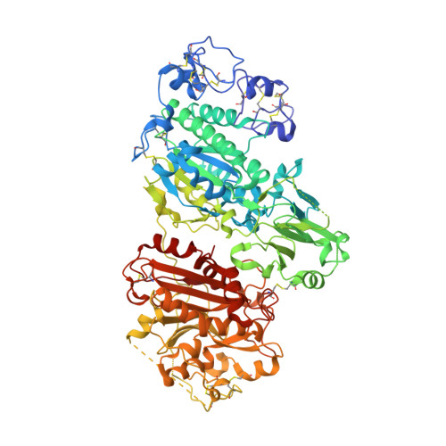
    7P0K

  • PubMed Abstract: 

    Autotaxin (ATX) is a secreted phosphodiesterase that has been implicated in a remarkably wide array of pathologies, especially in fibrosis and cancer. While ATX inhibitors have entered the clinical arena, a validated probe for positron emission tomography (PET) is currently lacking. With the aim to develop a suitable ATX-targeted PET radioligand, we have synthesized a focused library of fluorinated imidazo[1,2- a ]pyridine derivatives, determined their inhibition constants, and confirmed their binding mode by crystallographic analysis. Based on their promising in vitro properties, compounds 9c , 9f , 9h , and 9j were radiofluorinated. Also, a deuterated analog of [ 18 F] 9j , designated as [ 18 F]ATX-1905 ([ 18 F] 20 ), was designed and proved to be highly stable against in vivo radiodefluorination compared with [ 18 F] 9c , [ 18 F] 9f , [ 18 F] 9h , and [ 18 F] 9j . These results along with in vitro and in vivo studies toward ATX in a mouse model of LPS-induced liver injury suggest that [ 18 F]ATX-1905 is a suitable PET probe for the non-invasive quantification of ATX.


  • Organizational Affiliation
    • Division of Nuclear Medicine and Molecular Imaging, Massachusetts General Hospital & Department of Radiology, Harvard Medical School, Boston, Massachusetts 02114, United States.

Macromolecule Content 

  • Total Structure Weight: 91.6 kDa 
  • Atom Count: 6,691 
  • Modeled Residue Count: 770 
  • Deposited Residue Count: 770 
  • Unique protein chains: 1

Macromolecules

Find similar proteins by:|  3D Structure
Entity ID: 1
MoleculeChains  Sequence LengthOrganismDetailsImage
Isoform 2 of Ectonucleotide pyrophosphatase/phosphodiesterase family member 2A [auth AAA]770Rattus norvegicusMutation(s): 1 
Gene Names: Enpp2AtxNpps2
EC: 3.1.4.39 (PDB Primary Data), 3.1.4.4 (UniProt)
UniProt
Find proteins for Q64610 (Rattus norvegicus)
Explore Q64610 
Go to UniProtKB:  Q64610
Entity Groups
Sequence Clusters30% Identity50% Identity70% Identity90% Identity95% Identity100% Identity
UniProt GroupQ64610
Glycosylation
Glycosylation Sites: 1Go to GlyGen: Q64610-1
Sequence Annotations
Expand
Reference Sequence

Oligosaccharides

Help  
Entity ID: 2
MoleculeChains Length2D Diagram GlycosylationD Interactions
alpha-D-mannopyranose-(1-2)-alpha-D-mannopyranose-(1-3)-[alpha-D-mannopyranose-(1-6)]alpha-D-mannopyranose-(1-6)-[alpha-D-mannopyranose-(1-3)]beta-D-mannopyranose-(1-4)-2-acetamido-2-deoxy-beta-D-glucopyranoseB [auth AbA]7N/A
Glycosylation Resources
GlyTouCan: G19138OU
GlyCosmos: G19138OU
GlyGen: G19138OU

Small Molecules

Ligands 6 Unique
IDChains Name / Formula / InChI Key2D Diagram3D Interactions
4I0
(Subject of Investigation/LOI)

Query on 4I0


Download:Ideal Coordinates CCD File
L [auth AAA]2-[[2-ethyl-6-[4-[2-[(3~{R})-3-fluoranylpyrrolidin-1-yl]-2-oxidanylidene-ethyl]piperazin-1-yl]imidazo[1,2-a]pyridin-3-yl]-methyl-amino]-4-(4-fluorophenyl)-2,3-dihydro-1,3-thiazole-5-carbonitrile
C30 H34 F2 N8 O S
HIEJMXWIPDOGLP-RCRUUEGKSA-N
NAG

Query on NAG


Download:Ideal Coordinates CCD File
C [auth AAA]2-acetamido-2-deoxy-beta-D-glucopyranose
C8 H15 N O6
OVRNDRQMDRJTHS-FMDGEEDCSA-N
IOD

Query on IOD


Download:Ideal Coordinates CCD File
I [auth AAA],
J [auth AAA],
K [auth AAA],
M [auth AAA],
N [auth AAA]
IODIDE ION
I
XMBWDFGMSWQBCA-UHFFFAOYSA-M
ZN

Query on ZN


Download:Ideal Coordinates CCD File
F [auth AAA],
G [auth AAA]
ZINC ION
Zn
PTFCDOFLOPIGGS-UHFFFAOYSA-N
SCN

Query on SCN


Download:Ideal Coordinates CCD File
D [auth AAA],
E [auth AAA],
O [auth AAA]
THIOCYANATE ION
C N S
ZMZDMBWJUHKJPS-UHFFFAOYSA-M
CA

Query on CA


Download:Ideal Coordinates CCD File
H [auth AAA]CALCIUM ION
Ca
BHPQYMZQTOCNFJ-UHFFFAOYSA-N

Experimental Data & Validation

Experimental Data

  • Method: X-RAY DIFFRACTION
  • Resolution: 2.20 Å
  • R-Value Free:  0.271 (Depositor), 0.289 (DCC) 
  • R-Value Work:  0.212 (Depositor) 
Space Group: P 21 21 21
Unit Cell:
Length ( Å )Angle ( ˚ )
a = 61.57α = 90
b = 87.149β = 90
c = 145.361γ = 90
Software Package:
Software NamePurpose
REFMACrefinement
REFMACrefinement
Cootmodel building
XDSdata reduction
Aimlessdata scaling
MOLREPphasing

Structure Validation

View Full Validation Report


Ligand Structure Quality Assessment 


Entry History 

& Funding Information

Deposition Data


Funding OrganizationLocationGrant Number
Oncode InstituteNetherlands--

Revision History  (Full details and data files)

  • Version 1.0: 2022-07-13
    Type: Initial release
  • Version 1.1: 2024-01-31
    Changes: Data collection, Refinement description
  • Version 1.2: 2024-10-16
    Changes: Structure summary