


Crystal structure of IpgC in complex with a follow-up compound based on J20
Gardonyi, M., Heine, A., Klebe, G.To be published.
Experimental Data Snapshot
Starting Model: experimental
View more details
Entity ID: 1 | |||||
|---|---|---|---|---|---|
| Molecule | Chains | Sequence Length | Organism | Details | Image |
| Chaperone protein IpgC | 143 | Shigella flexneri | Mutation(s): 0 Gene Names: ipgC, ippI, CP0129 | 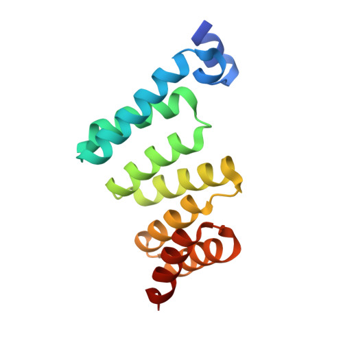 | |
UniProt | |||||
Entity Groups | |||||
| Sequence Clusters | 30% Identity50% Identity70% Identity90% Identity95% Identity100% Identity | ||||
| UniProt Group | P0A2U4 | ||||
Sequence AnnotationsExpand | |||||
Reference Sequence | |||||
| Ligands 4 Unique | |||||
|---|---|---|---|---|---|
| ID | Chains | Name / Formula / InChI Key | 2D Diagram | 3D Interactions | |
| 2IY Download:Ideal Coordinates CCD File | E [auth B] | 3-(4-chlorophenyl)-5-methylsulfanyl-1~{H}-pyrazole-4-carbonitrile C11 H8 Cl N3 S KRQSUBRVYHNKPM-UHFFFAOYSA-N |  | ||
| PGE Download:Ideal Coordinates CCD File | G [auth B] | TRIETHYLENE GLYCOL C6 H14 O4 ZIBGPFATKBEMQZ-UHFFFAOYSA-N |  | ||
| DMS Download:Ideal Coordinates CCD File | C [auth A] | DIMETHYL SULFOXIDE C2 H6 O S IAZDPXIOMUYVGZ-UHFFFAOYSA-N |  | ||
| CL Download:Ideal Coordinates CCD File | D [auth A], F [auth B] | CHLORIDE ION Cl VEXZGXHMUGYJMC-UHFFFAOYSA-M |  | ||
| Length ( Å ) | Angle ( ˚ ) |
|---|---|
| a = 57.788 | α = 90 |
| b = 57.788 | β = 90 |
| c = 160.301 | γ = 120 |
| Software Name | Purpose |
|---|---|
| PHENIX | refinement |
| XDS | data reduction |
| XDS | data scaling |
| PHASER | phasing |
| Coot | model building |


