7OTE | pdb_00007ote

Src Kinase Domain in complex with ponatinib


Experimental Data Snapshot

  • Method: X-RAY DIFFRACTION
  • Resolution: 2.49 Å
  • R-Value Free: 
    0.252 (Depositor), 0.253 (DCC) 
  • R-Value Work: 
    0.175 (Depositor), 0.188 (DCC) 
  • R-Value Observed: 
    0.179 (Depositor) 

Starting Model: experimental
View more details

wwPDB Validation 3D Report Full Report

Validation slider image for 7OTE

Ligand Structure Quality Assessment 


This is version 1.3 of the entry. See complete history

Literature

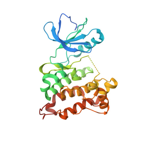
An allosteric switch between the activation loop and a c-terminal palindromic phospho-motif controls c-Src function.

Cuesta-Hernandez, H.N.Contreras, J.Soriano-Maldonado, P.Sanchez-Wandelmer, J.Yeung, W.Martin-Hurtado, A.Munoz, I.G.Kannan, N.Llimargas, M.Munoz, J.Plaza-Menacho, I.

(2023) Nat Commun 14: 6548-6548

  • DOI: https://doi.org/10.1038/s41467-023-41890-7
  • Primary Citation Related Structures: 
    7OTE

  • PubMed Abstract: 

    Autophosphorylation controls the transition between discrete functional and conformational states in protein kinases, yet the structural and molecular determinants underlying this fundamental process remain unclear. Here we show that c-terminal Tyr 530 is a de facto c-Src autophosphorylation site with slow time-resolution kinetics and a strong intermolecular component. On the contrary, activation-loop Tyr 419 undergoes faster kinetics and a cis-to-trans phosphorylation switch that controls c-terminal Tyr 530 autophosphorylation, enzyme specificity, and strikingly, c-Src non-catalytic function as a substrate. In line with this, we visualize by X-ray crystallography a snapshot of Tyr 530 intermolecular autophosphorylation. In an asymmetric arrangement of both catalytic domains, a c-terminal palindromic phospho-motif flanking Tyr 530 on the substrate molecule engages the G-loop of the active kinase adopting a position ready for entry into the catalytic cleft. Perturbation of the phospho-motif accounts for c-Src dysfunction as indicated by viral and colorectal cancer (CRC)-associated c-terminal deleted variants. We show that c-terminal residues 531 to 536 are required for c-Src Tyr 530 autophosphorylation, and such a detrimental effect is caused by the substrate molecule inhibiting allosterically the active kinase. Our work reveals a crosstalk between the activation and c-terminal segments that control the allosteric interplay between substrate- and enzyme-acting kinases during autophosphorylation.


  • Organizational Affiliation
    • Kinases, Protein Phosphorylation and Cancer Group, Structural Biology Programme, Spanish National Cancer Research Center (CNIO), C/Melchor Fernández Almagro num. 3, 28029, Madrid, Spain.

Macromolecule Content 

  • Total Structure Weight: 69.28 kDa 
  • Atom Count: 4,388 
  • Modeled Residue Count: 518 
  • Deposited Residue Count: 586 
  • Unique protein chains: 1

Macromolecules

Find similar proteins by:|  3D Structure
Entity ID: 1
MoleculeChains  Sequence LengthOrganismDetailsImage
Proto-oncogene tyrosine-protein kinase Src
A, B
293Homo sapiensMutation(s): 0 
Gene Names: SRCSRC1
EC: 2.7.10.2
UniProt & NIH Common Fund Data Resources
Find proteins for P12931 (Homo sapiens)
Explore P12931 
Go to UniProtKB:  P12931
PHAROS:  P12931
GTEx:  ENSG00000197122 
Entity Groups
Sequence Clusters30% Identity50% Identity70% Identity90% Identity95% Identity100% Identity
UniProt GroupP12931
Sequence Annotations
Expand
Reference Sequence

Small Molecules

Ligands 3 Unique
IDChains Name / Formula / InChI Key2D Diagram3D Interactions
0LI
(Subject of Investigation/LOI)

Query on 0LI


Download:Ideal Coordinates CCD File
C [auth A],
I [auth B]
3-(imidazo[1,2-b]pyridazin-3-ylethynyl)-4-methyl-N-{4-[(4-methylpiperazin-1-yl)methyl]-3-(trifluoromethyl)phenyl}benzam ide
C29 H27 F3 N6 O
PHXJVRSECIGDHY-UHFFFAOYSA-N
TRS

Query on TRS


Download:Ideal Coordinates CCD File
D [auth A]2-AMINO-2-HYDROXYMETHYL-PROPANE-1,3-DIOL
C4 H12 N O3
LENZDBCJOHFCAS-UHFFFAOYSA-O
GOL

Query on GOL


Download:Ideal Coordinates CCD File
E [auth A]
F [auth A]
G [auth A]
H [auth A]
J [auth B]
E [auth A],
F [auth A],
G [auth A],
H [auth A],
J [auth B],
K [auth B],
L [auth B],
M [auth B],
N [auth B],
O [auth B]
GLYCEROL
C3 H8 O3
PEDCQBHIVMGVHV-UHFFFAOYSA-N
Binding Affinity Annotations 
IDSourceBinding Affinity
0LI BindingDB:  7OTE Kd: 0.7 (nM) from 1 assay(s)
IC50: min: 0.89, max: 10 (nM) from 6 assay(s)

Experimental Data & Validation

Experimental Data

  • Method: X-RAY DIFFRACTION
  • Resolution: 2.49 Å
  • R-Value Free:  0.252 (Depositor), 0.253 (DCC) 
  • R-Value Work:  0.175 (Depositor), 0.188 (DCC) 
  • R-Value Observed: 0.179 (Depositor) 
Space Group: P 1 21 1
Unit Cell:
Length ( Å )Angle ( ˚ )
a = 42.153α = 90
b = 124.631β = 90.15
c = 63.591γ = 90
Software Package:
Software NamePurpose
REFMACrefinement
MOSFLMdata reduction
Aimlessdata scaling
PHASERphasing
PDB_EXTRACTdata extraction

Structure Validation

View Full Validation Report


Ligand Structure Quality Assessment 


Entry History 

& Funding Information

Deposition Data


Funding OrganizationLocationGrant Number
Spanish Ministry of Science, Innovation, and UniversitiesSpainBFU2017-86710-R
Spanish Ministry of Science, Innovation, and UniversitiesSpainRYC-2016-1938

Revision History  (Full details and data files)

  • Version 1.0: 2022-06-22
    Type: Initial release
  • Version 1.1: 2023-08-02
    Changes: Database references
  • Version 1.2: 2024-01-31
    Changes: Data collection, Refinement description
  • Version 1.3: 2024-03-27
    Changes: Database references