7OTB | pdb_00007otb

Ruthenium polypridyl complex bound to a unimolecular chair-form G-quadruplex


Experimental Data Snapshot

  • Method: X-RAY DIFFRACTION
  • Resolution: 1.60 Å
  • R-Value Free: 
    0.181 (Depositor), 0.190 (DCC) 
  • R-Value Work: 
    0.165 (Depositor), 0.180 (DCC) 
  • R-Value Observed: 
    0.166 (Depositor) 

wwPDB Validation   3D Report Full Report


Ligand Structure Quality Assessment 


This is version 1.2 of the entry. See complete history


Literature

Ruthenium Polypyridyl Complex Bound to a Unimolecular Chair-Form G-Quadruplex.

McQuaid, K.T.Takahashi, S.Baumgaertner, L.Cardin, D.J.Paterson, N.G.Hall, J.P.Sugimoto, N.Cardin, C.J.

(2022) J Am Chem Soc 144: 5956-5964

  • DOI: https://doi.org/10.1021/jacs.2c00178
  • Primary Citation of Related Structures:  
    7OTB

  • PubMed Abstract: 

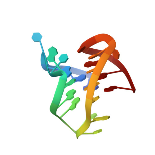
    The DNA G-quadruplex is known for forming a range of topologies and for the observed lability of the assembly, consistent with its transient formation in live cells. The stabilization of a particular topology by a small molecule is of great importance for therapeutic applications. Here, we show that the ruthenium complex Λ-[Ru(phen) 2 (qdppz)] 2+ displays enantiospecific G-quadruplex binding. It crystallized in 1:1 stoichiometry with a modified human telomeric G-quadruplex sequence, GGGTTAGGGTTAGGGTTTGGG ( htel21 T 18 ), in an antiparallel chair topology, the first structurally characterized example of ligand binding to this topology. The lambda complex is bound in an intercalation cavity created by a terminal G-quartet and the central narrow lateral loop formed by T 10 -T 11 -A 12 . The two remaining wide lateral loops are linked through a third K + ion at the other end of the G-quartet stack, which also coordinates three thymine residues. In a comparative ligand-binding study, we showed, using a Klenow fragment assay, that this complex is the strongest observed inhibitor of replication, both using the native human telomeric sequence and the modified sequence used in this work.


  • Organizational Affiliation
    • Department of Chemistry, University of Reading, Whiteknights, Reading RG6 6AD, U.K.

Macromolecules

Find similar nucleic acids by:  Sequence   |   3D Structure  

Entity ID: 1
MoleculeChains LengthOrganismImage
DNA (5'-D(*GP*GP*GP*TP*TP*AP*GP*GP*GP*TP*TP*AP*GP*GP*GP*TP*TP*TP*GP*GP*G)-3')21synthetic construct
Sequence Annotations
Expand
  • Reference Sequence
Small Molecules
Ligands 3 Unique
IDChains Name / Formula / InChI Key2D Diagram3D Interactions
0K8 (Subject of Investigation/LOI)
Query on 0K8

Download Ideal Coordinates CCD File 
G [auth A]Ruthenium bis-(phenanthroline) 12,17-dihydro-naphthodipyridophenazine-12,17-dione
C50 H28 N8 O2 Ru
LEJPQQFNZLHEQD-UHFFFAOYSA-N
BA
Query on BA

Download Ideal Coordinates CCD File 
F [auth A]BARIUM ION
Ba
XDFCIPNJCBUZJN-UHFFFAOYSA-N
K
Query on K

Download Ideal Coordinates CCD File 
B [auth A],
C [auth A],
D [auth A],
E [auth A]
POTASSIUM ION
K
NPYPAHLBTDXSSS-UHFFFAOYSA-N
Experimental Data & Validation

Experimental Data

  • Method: X-RAY DIFFRACTION
  • Resolution: 1.60 Å
  • R-Value Free:  0.181 (Depositor), 0.190 (DCC) 
  • R-Value Work:  0.165 (Depositor), 0.180 (DCC) 
  • R-Value Observed: 0.166 (Depositor) 
Space Group: P 32 2 1
Unit Cell:
Length ( Å )Angle ( ˚ )
a = 29.68α = 90
b = 29.68β = 90
c = 113.98γ = 120
Software Package:
Software NamePurpose
PHENIXrefinement
PDB_EXTRACTdata extraction
xia2data reduction
XDSdata reduction
xia2data scaling
XSCALEdata scaling
SHELXDEphasing
Cootmodel building

Structure Validation

View Full Validation Report


Ligand Structure Quality Assessment 


Entry History & Funding Information

Deposition Data


Funding OrganizationLocationGrant Number
Biotechnology and Biological Sciences Research Council (BBSRC)United KingdomBB/T008342/1

Revision History  (Full details and data files)

  • Version 1.0: 2022-04-06
    Type: Initial release
  • Version 1.1: 2022-04-20
    Changes: Database references
  • Version 1.2: 2024-06-19
    Changes: Data collection