7OP3 | pdb_00007op3

Cryo-EM structure of P5B-ATPase E2PiSPM


Experimental Data Snapshot

  • Method: ELECTRON MICROSCOPY
  • Resolution: 3.50 Å
  • Aggregation State: PARTICLE 
  • Reconstruction Method: SINGLE PARTICLE 

Starting Model: experimental
View more details

wwPDB Validation 3D Report Full Report

Validation slider image for 7OP3

This is version 1.2 of the entry. See complete history

Literature

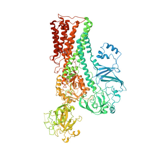
Structure and transport mechanism of P5B-ATPases.

Li, P.Wang, K.Salustros, N.Gronberg, C.Gourdon, P.

(2021) Nat Commun 12: 3973-3973

  • DOI: https://doi.org/10.1038/s41467-021-24148-y
  • Primary Citation Related Structures: 
    7OP1, 7OP3, 7OP5, 7OP8

  • PubMed Abstract: 

    In human cells, P5B-ATPases execute the active export of physiologically important polyamines such as spermine from lysosomes to the cytosol, a function linked to a palette of disorders. Yet, the overall shape of P5B-ATPases and the mechanisms of polyamine recognition, uptake and transport remain elusive. Here we describe a series of cryo-electron microscopy structures of a yeast homolog of human ATP13A2-5, Ypk9, determined at resolutions reaching 3.4 Å, and depicting three separate transport cycle intermediates, including spermine-bound conformations. Surprisingly, in the absence of cargo, Ypk9 rests in a phosphorylated conformation auto-inhibited by the N-terminus. Spermine uptake is accomplished through an electronegative cleft lined by transmembrane segments 2, 4 and 6. Despite the dramatically different nature of the transported cargo, these findings pinpoint shared principles of transport and regulation among the evolutionary related P4-, P5A- and P5B-ATPases. The data also provide a framework for analysis of associated maladies, such as Parkinson's disease.


  • Organizational Affiliation
    • Department of Experimental Medical Science, Lund University, Lund, Sweden.

Macromolecule Content 

  • Total Structure Weight: 156.34 kDa 
  • Atom Count: 8,772 
  • Modeled Residue Count: 1,100 
  • Deposited Residue Count: 1,394 
  • Unique protein chains: 1

Macromolecules

Find similar proteins by:|  3D Structure
Entity ID: 1
MoleculeChains  Sequence LengthOrganismDetailsImage
Cation-transporting ATPase1,394Thermochaetoides thermophila DSM 1495Mutation(s): 0 
EC: 7.2.2
Membrane Entity: Yes 
UniProt
Find proteins for G0S7G9 (Chaetomium thermophilum (strain DSM 1495 / CBS 144.50 / IMI 039719))
Explore G0S7G9 
Go to UniProtKB:  G0S7G9
Entity Groups
Sequence Clusters30% Identity50% Identity70% Identity90% Identity95% Identity100% Identity
UniProt GroupG0S7G9
Sequence Annotations
Expand
Reference Sequence

Small Molecules

Ligands 1 Unique
IDChains Name / Formula / InChI Key2D Diagram3D Interactions
SPM
(Subject of Investigation/LOI)

Query on SPM


Download:Ideal Coordinates CCD File
B [auth A]SPERMINE
C10 H26 N4
PFNFFQXMRSDOHW-UHFFFAOYSA-N

Experimental Data & Validation

Experimental Data

  • Method: ELECTRON MICROSCOPY
  • Resolution: 3.50 Å
  • Aggregation State: PARTICLE 
  • Reconstruction Method: SINGLE PARTICLE 
EM Software:
TaskSoftware PackageVersion
MODEL REFINEMENTPHENIX
MODEL REFINEMENTCoot

Structure Validation

View Full Validation Report


Entry History 

& Funding Information

Deposition Data


Funding OrganizationLocationGrant Number
Knut and Alice Wallenberg FoundationSweden2020.0914, 2015.0313
LundbeckfondenSwedenR313-2019-774, R218-2016-1548, R133-A12689

Revision History  (Full details and data files)

  • Version 1.0: 2021-06-30
    Type: Initial release
  • Version 1.1: 2021-07-07
    Changes: Database references
  • Version 1.2: 2024-05-01
    Changes: Data collection, Database references, Refinement description