7O9L

Dictyostelium discoideum dye decolorizing peroxidase DyPA in complex with cyanide.


Experimental Data Snapshot

  • Method: X-RAY DIFFRACTION
  • Resolution: 1.85 Å
  • R-Value Free: 0.180 
  • R-Value Work: 0.155 
  • R-Value Observed: 0.156 

wwPDB Validation   3D Report Full Report


Ligand Structure Quality Assessment 


This is version 1.1 of the entry. See complete history


Literature

Structural and Biochemical Characterization of a Dye-Decolorizing Peroxidase from Dictyostelium discoideum .

Rai, A.Klare, J.P.Reinke, P.Y.A.Englmaier, F.Fohrer, J.Fedorov, R.Taft, M.H.Chizhov, I.Curth, U.Plettenburg, O.Manstein, D.J.

(2021) Int J Mol Sci 22

  • DOI: https://doi.org/10.3390/ijms22126265
  • Primary Citation of Related Structures:  
    7O9J, 7O9L, 7ODZ

  • PubMed Abstract: 

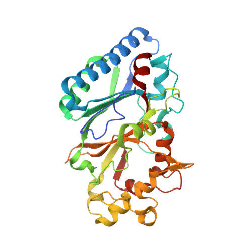
    A novel cytoplasmic dye-decolorizing peroxidase from Dictyostelium discoideum was investigated that oxidizes anthraquinone dyes, lignin model compounds, and general peroxidase substrates such as ABTS efficiently. Unlike related enzymes, an aspartate residue replaces the first glycine of the conserved GXXDG motif in Dictyostelium DyPA. In solution, Dictyostelium DyPA exists as a stable dimer with the side chain of Asp146 contributing to the stabilization of the dimer interface by extending the hydrogen bond network connecting two monomers. To gain mechanistic insights, we solved the Dictyostelium DyPA structures in the absence of substrate as well as in the presence of potassium cyanide and veratryl alcohol to 1.7, 1.85, and 1.6 Å resolution, respectively. The active site of Dictyostelium DyPA has a hexa-coordinated heme iron with a histidine residue at the proximal axial position and either an activated oxygen or CN - molecule at the distal axial position. Asp149 is in an optimal conformation to accept a proton from H 2 O 2 during the formation of compound I. Two potential distal solvent channels and a conserved shallow pocket leading to the heme molecule were found in Dictyostelium DyPA. Further, we identified two substrate-binding pockets per monomer in Dictyostelium DyPA at the dimer interface. Long-range electron transfer pathways associated with a hydrogen-bonding network that connects the substrate-binding sites with the heme moiety are described.


  • Organizational Affiliation

    Institute for Biophysical Chemistry, Hannover Medical School, Fritz Hartmann Centre for Medical Research Carl Neuberg Str. 1, D-30625 Hannover, Germany.


Macromolecules
Find similar proteins by:  (by identity cutoff)  |  3D Structure
Entity ID: 1
MoleculeChains Sequence LengthOrganismDetailsImage
DyPA
A, B
311Dictyostelium discoideumMutation(s): 0 
Gene Names: DDB0168077DDB0217308
UniProt
Find proteins for Q556V8 (Dictyostelium discoideum)
Explore Q556V8 
Go to UniProtKB:  Q556V8
Entity Groups  
Sequence Clusters30% Identity50% Identity70% Identity90% Identity95% Identity100% Identity
UniProt GroupQ556V8
Sequence Annotations
Expand
  • Reference Sequence
Small Molecules
Ligands 3 Unique
IDChains Name / Formula / InChI Key2D Diagram3D Interactions
HEM (Subject of Investigation/LOI)
Query on HEM

Download Ideal Coordinates CCD File 
C [auth A],
I [auth B]
PROTOPORPHYRIN IX CONTAINING FE
C34 H32 Fe N4 O4
KABFMIBPWCXCRK-RGGAHWMASA-L
EDO
Query on EDO

Download Ideal Coordinates CCD File 
D [auth A]
E [auth A]
F [auth A]
G [auth A]
J [auth B]
D [auth A],
E [auth A],
F [auth A],
G [auth A],
J [auth B],
K [auth B],
L [auth B]
1,2-ETHANEDIOL
C2 H6 O2
LYCAIKOWRPUZTN-UHFFFAOYSA-N
CYN (Subject of Investigation/LOI)
Query on CYN

Download Ideal Coordinates CCD File 
H [auth A],
M [auth B]
CYANIDE ION
C N
XFXPMWWXUTWYJX-UHFFFAOYSA-N
Experimental Data & Validation

Experimental Data

  • Method: X-RAY DIFFRACTION
  • Resolution: 1.85 Å
  • R-Value Free: 0.180 
  • R-Value Work: 0.155 
  • R-Value Observed: 0.156 
  • Space Group: P 41 21 2
Unit Cell:
Length ( Å )Angle ( ˚ )
a = 141.05α = 90
b = 141.05β = 90
c = 95.48γ = 90
Software Package:
Software NamePurpose
PHENIXrefinement
PHENIXrefinement
XDSdata reduction
SADABSdata scaling
PHASERphasing

Structure Validation

View Full Validation Report


Ligand Structure Quality Assessment 


Entry History & Funding Information

Deposition Data


Funding OrganizationLocationGrant Number
German Research Foundation (DFG)GermanyEXC 2155 - 39087428-B11

Revision History  (Full details and data files)

  • Version 1.0: 2021-07-14
    Type: Initial release
  • Version 1.1: 2024-01-31
    Changes: Data collection, Database references, Refinement description