7O7G | pdb_00007o7g

Crystal structure of the Shewanella oneidensis MR1 MtrC mutant H561M


Experimental Data Snapshot

  • Method: X-RAY DIFFRACTION
  • Resolution: 1.60 Å
  • R-Value Free: 
    0.205 (Depositor), 0.214 (DCC) 
  • R-Value Work: 
    0.170 (Depositor), 0.181 (DCC) 
  • R-Value Observed: 
    0.172 (Depositor) 

Starting Model: experimental
View more details

wwPDB Validation 3D Report Full Report

Validation slider image for 7O7G

Ligand Structure Quality Assessment 


This is version 1.2 of the entry. See complete history

Literature

Nanosecond heme-to-heme electron transfer rates in a multiheme cytochrome nanowire reported by a spectrally unique His/Met-ligated heme.

van Wonderen, J.H.Adamczyk, K.Wu, X.Jiang, X.Piper, S.E.H.Hall, C.R.Edwards, M.J.Clarke, T.A.Zhang, H.Jeuken, L.J.C.Sazanovich, I.V.Towrie, M.Blumberger, J.Meech, S.R.Butt, J.N.

(2021) Proc Natl Acad Sci U S A 118

  • DOI: https://doi.org/10.1073/pnas.2107939118
  • Primary Citation Related Structures: 
    7O7G

  • PubMed Abstract: 

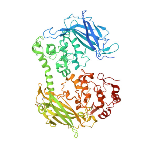
    Proteins achieve efficient energy storage and conversion through electron transfer along a series of redox cofactors. Multiheme cytochromes are notable examples. These proteins transfer electrons over distance scales of several nanometers to >10 μm and in so doing they couple cellular metabolism with extracellular redox partners including electrodes. Here, we report pump-probe spectroscopy that provides a direct measure of the intrinsic rates of heme-heme electron transfer in this fascinating class of proteins. Our study took advantage of a spectrally unique His/Met-ligated heme introduced at a defined site within the decaheme extracellular MtrC protein of Shewanella oneidensis We observed rates of heme-to-heme electron transfer on the order of 10 9 s -1 (3.7 to 4.3 Å edge-to-edge distance), in good agreement with predictions based on density functional and molecular dynamics calculations. These rates are among the highest reported for ground-state electron transfer in biology. Yet, some fall 2 to 3 orders of magnitude below the Moser-Dutton ruler because electron transfer at these short distances is through space and therefore associated with a higher tunneling barrier than the through-protein tunneling scenario that is usual at longer distances. Moreover, we show that the His/Met-ligated heme creates an electron sink that stabilizes the charge separated state on the 100-μs time scale. This feature could be exploited in future designs of multiheme cytochromes as components of versatile photosynthetic biohybrid assemblies.


  • Organizational Affiliation
    • School of Chemistry, University of East Anglia, Norwich Research Park, Norwich NR4 7TJ, United Kingdom.

Macromolecule Content 

  • Total Structure Weight: 78.45 kDa 
  • Atom Count: 6,065 
  • Modeled Residue Count: 627 
  • Deposited Residue Count: 671 
  • Unique protein chains: 1

Macromolecules

Find similar proteins by:|  3D Structure
Entity ID: 1
MoleculeChains  Sequence LengthOrganismDetailsImage
Extracellular iron oxide respiratory system surface decaheme cytochrome c component MtrC671Shewanella oneidensis MR-1Mutation(s): 1 
Gene Names: mtrCSO_1778
UniProt
Find proteins for Q8EG34 (Shewanella oneidensis (strain ATCC 700550 / JCM 31522 / CIP 106686 / LMG 19005 / NCIMB 14063 / MR-1))
Explore Q8EG34 
Go to UniProtKB:  Q8EG34
Entity Groups
Sequence Clusters30% Identity50% Identity70% Identity90% Identity95% Identity100% Identity
UniProt GroupQ8EG34
Sequence Annotations
Expand
Reference Sequence

Small Molecules

Ligands 4 Unique
IDChains Name / Formula / InChI Key2D Diagram3D Interactions
HEC

Query on HEC


Download:Ideal Coordinates CCD File
B [auth A]
C [auth A]
D [auth A]
E [auth A]
F [auth A]
B [auth A],
C [auth A],
D [auth A],
E [auth A],
F [auth A],
G [auth A],
H [auth A],
I [auth A],
J [auth A],
K [auth A]
HEME C
C34 H34 Fe N4 O4
HXQIYSLZKNYNMH-LJNAALQVSA-N
EDO

Query on EDO


Download:Ideal Coordinates CCD File
BA [auth A]
P [auth A]
Q [auth A]
R [auth A]
S [auth A]
BA [auth A],
P [auth A],
Q [auth A],
R [auth A],
S [auth A],
T [auth A],
U [auth A],
V [auth A]
1,2-ETHANEDIOL
C2 H6 O2
LYCAIKOWRPUZTN-UHFFFAOYSA-N
ACT

Query on ACT


Download:Ideal Coordinates CCD File
AA [auth A],
W [auth A],
X [auth A],
Y [auth A],
Z [auth A]
ACETATE ION
C2 H3 O2
QTBSBXVTEAMEQO-UHFFFAOYSA-M
CA

Query on CA


Download:Ideal Coordinates CCD File
L [auth A],
M [auth A],
N [auth A],
O [auth A]
CALCIUM ION
Ca
BHPQYMZQTOCNFJ-UHFFFAOYSA-N

Experimental Data & Validation

Experimental Data

  • Method: X-RAY DIFFRACTION
  • Resolution: 1.60 Å
  • R-Value Free:  0.205 (Depositor), 0.214 (DCC) 
  • R-Value Work:  0.170 (Depositor), 0.181 (DCC) 
  • R-Value Observed: 0.172 (Depositor) 
Space Group: P 21 21 21
Unit Cell:
Length ( Å )Angle ( ˚ )
a = 52.875α = 90
b = 89.657β = 90
c = 153.9γ = 90
Software Package:
Software NamePurpose
REFMACrefinement
PDB_EXTRACTdata extraction
DIALSdata reduction
DIALSdata scaling
PHASERphasing

Structure Validation

View Full Validation Report


Ligand Structure Quality Assessment 


Entry History 

& Funding Information

Deposition Data


Funding OrganizationLocationGrant Number
Biotechnology and Biological Sciences Research Council (BBSRC)United KingdomBB/P01819X/1
Biotechnology and Biological Sciences Research Council (BBSRC)United KingdomBB/S002499/1
Engineering and Physical Sciences Research CouncilUnited KingdomEP/M001989/1

Revision History  (Full details and data files)

  • Version 1.0: 2021-10-06
    Type: Initial release
  • Version 1.1: 2024-01-31
    Changes: Data collection, Refinement description
  • Version 1.2: 2024-11-13
    Changes: Structure summary