7O76 | pdb_00007o76

Reversible supramolecular assembly of the anti-microbial peptide plectasin


Experimental Data Snapshot

  • Method: X-RAY DIFFRACTION
  • Resolution: 1.13 Å
  • R-Value Free: 
    0.178 (Depositor), 0.179 (DCC) 
  • R-Value Work: 
    0.146 (Depositor), 0.143 (DCC) 

Starting Model: experimental
View more details

wwPDB Validation 3D Report Full Report

Validation slider image for 7O76

This is version 1.2 of the entry. See complete history

Literature

pH- and concentration-dependent supramolecular assembly of a fungal defensin plectasin variant into helical non-amyloid fibrils.

Pohl, C.Effantin, G.Kandiah, E.Meier, S.Zeng, G.Streicher, W.Segura, D.R.Mygind, P.H.Sandvang, D.Nielsen, L.A.Peters, G.H.J.Schoehn, G.Mueller-Dieckmann, C.Noergaard, A.Harris, P.

(2022) Nat Commun 13: 3162-3162

  • DOI: https://doi.org/10.1038/s41467-022-30462-w
  • Primary Citation Related Structures: 
    7O76, 7OAE, 7OAG

  • PubMed Abstract: 

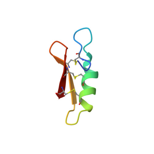
    Self-assembly and fibril formation play important roles in protein behaviour. Amyloid fibril formation is well-studied due to its role in neurodegenerative diseases and characterized by refolding of the protein into predominantly β-sheet form. However, much less is known about the assembly of proteins into other types of supramolecular structures. Using cryo-electron microscopy at a resolution of 1.97 Å, we show that a triple-mutant of the anti-microbial peptide plectasin, PPI42, assembles into helical non-amyloid fibrils. The in vitro anti-microbial activity was determined and shown to be enhanced compared to the wildtype. Plectasin contains a cysteine-stabilised α-helix-β-sheet structure, which remains intact upon fibril formation. Two protofilaments form a right-handed protein fibril. The fibril formation is reversible and follows sigmoidal kinetics with a pH- and concentration dependent equilibrium between soluble monomer and protein fibril. This high-resolution structure reveals that α/β proteins can natively assemble into fibrils.


  • Organizational Affiliation
    • Novozymes A/S, Bagsvaerd, Denmark. christin.pohl@biochemistry.lu.se.

Macromolecule Content 

  • Total Structure Weight: 4.52 kDa 
  • Atom Count: 352 
  • Modeled Residue Count: 40 
  • Deposited Residue Count: 40 
  • Unique protein chains: 1

Macromolecules

Find similar proteins by:|  3D Structure
Entity ID: 1
MoleculeChains  Sequence LengthOrganismDetailsImage
Fungal defensin plectasinA [auth XXX]40Pseudoplectania nigrellaMutation(s): 0 
Gene Names: DEF
UniProt
Find proteins for Q53I06 (Pseudoplectania nigrella)
Explore Q53I06 
Go to UniProtKB:  Q53I06
Entity Groups
Sequence Clusters30% Identity50% Identity70% Identity90% Identity95% Identity100% Identity
UniProt GroupQ53I06
Sequence Annotations
Expand
Reference Sequence

Small Molecules

Ligands 1 Unique
IDChains Name / Formula / InChI Key2D Diagram3D Interactions
PEG

Query on PEG


Download:Ideal Coordinates CCD File
B [auth XXX]DI(HYDROXYETHYL)ETHER
C4 H10 O3
MTHSVFCYNBDYFN-UHFFFAOYSA-N

Experimental Data & Validation

Experimental Data

  • Method: X-RAY DIFFRACTION
  • Resolution: 1.13 Å
  • R-Value Free:  0.178 (Depositor), 0.179 (DCC) 
  • R-Value Work:  0.146 (Depositor), 0.143 (DCC) 
Space Group: P 1 21 1
Unit Cell:
Length ( Å )Angle ( ˚ )
a = 23.678α = 90
b = 20.093β = 104.279
c = 32.546γ = 90
Software Package:
Software NamePurpose
REFMACrefinement
XDSdata reduction
XSCALEdata scaling
MOLREPphasing

Structure Validation

View Full Validation Report


Entry History 

& Funding Information

Deposition Data


Funding OrganizationLocationGrant Number
H2020 Marie Curie Actions of the European CommissionEuropean Union675074

Revision History  (Full details and data files)

  • Version 1.0: 2022-06-29
    Type: Initial release
  • Version 1.1: 2024-01-31
    Changes: Data collection, Refinement description
  • Version 1.2: 2024-11-20
    Changes: Structure summary