7O68

Crystal structure of human mitochondrial ferritin (hMTF) Fe(II)-loaded for 120 minutes showing either a dioxygen or a superoxide anion coordinated to iron ions in the ferroxidase site.


Experimental Data Snapshot

  • Method: X-RAY DIFFRACTION
  • Resolution: 1.68 Å
  • R-Value Free: 0.183 
  • R-Value Work: 0.158 
  • R-Value Observed: 0.159 

wwPDB Validation   3D Report Full Report


This is version 1.1 of the entry. See complete history


Literature

Iron Binding in the Ferroxidase Site of Human Mitochondrial Ferritin.

Ciambellotti, S.Pratesi, A.Tassone, G.Turano, P.Mangani, S.Pozzi, C.

(2021) Chemistry 27: 14690-14701

  • DOI: https://doi.org/10.1002/chem.202102270
  • Primary Citation of Related Structures:  
    7O63, 7O64, 7O65, 7O66, 7O67, 7O68, 7O69, 7O6A, 7O6C, 7O6D, 7OWY

  • PubMed Abstract: 

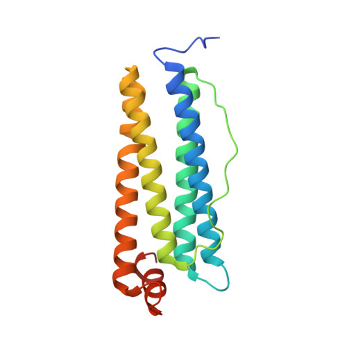
    Ferritins are nanocage proteins that store iron ions in their central cavity as hydrated ferric oxide biominerals. In mammals, further the L (light) and H (heavy) chains constituting cytoplasmic maxi-ferritins, an additional type of ferritin has been identified, the mitochondrial ferritin (MTF). Human MTF (hMTF) is a functional homopolymeric H-like ferritin performing the ferroxidase activity in its ferroxidase site (FS), in which Fe(II) is oxidized to Fe(III) in the presence of dioxygen. To better investigate its ferroxidase properties, here we performed time-lapse X-ray crystallography analysis of hMTF, providing structural evidence of how iron ions interact with hMTF and of their binding to the FS. Transient iron binding sites, populating the pathway along the cage from the iron entry channel to the catalytic center, were also identified. Furthermore, our kinetic data at variable iron loads indicate that the catalytic iron oxidation reaction occurs via a diferric peroxo intermediate followed by the formation of ferric-oxo species, with significant differences with respect to human H-type ferritin.


  • Organizational Affiliation

    Department of Chemistry "Ugo Schiff" Department of Excellence 2018-2022, University of Florence, via della Lastruccia 2, 50019, Sesto Fiorentino, Italy.


Macromolecules
Find similar proteins by:  (by identity cutoff)  |  3D Structure
Entity ID: 1
MoleculeChains Sequence LengthOrganismDetailsImage
Ferritin, mitochondrial183Homo sapiensMutation(s): 0 
Gene Names: FTMT
EC: 1.16.3.1
UniProt & NIH Common Fund Data Resources
Find proteins for Q8N4E7 (Homo sapiens)
Explore Q8N4E7 
Go to UniProtKB:  Q8N4E7
PHAROS:  Q8N4E7
GTEx:  ENSG00000181867 
Entity Groups  
Sequence Clusters30% Identity50% Identity70% Identity90% Identity95% Identity100% Identity
UniProt GroupQ8N4E7
Sequence Annotations
Expand
  • Reference Sequence
Small Molecules
Ligands 4 Unique
IDChains Name / Formula / InChI Key2D Diagram3D Interactions
FE2 (Subject of Investigation/LOI)
Query on FE2

Download Ideal Coordinates CCD File 
B [auth A]
C [auth A]
D [auth A]
E [auth A]
F [auth A]
B [auth A],
C [auth A],
D [auth A],
E [auth A],
F [auth A],
G [auth A],
H [auth A],
I [auth A],
J [auth A],
K [auth A],
L [auth A]
FE (II) ION
Fe
CWYNVVGOOAEACU-UHFFFAOYSA-N
CL
Query on CL

Download Ideal Coordinates CCD File 
N [auth A],
O [auth A],
P [auth A],
Q [auth A]
CHLORIDE ION
Cl
VEXZGXHMUGYJMC-UHFFFAOYSA-M
OXY
Query on OXY

Download Ideal Coordinates CCD File 
M [auth A]OXYGEN MOLECULE
O2
MYMOFIZGZYHOMD-UHFFFAOYSA-N
MG
Query on MG

Download Ideal Coordinates CCD File 
R [auth A]MAGNESIUM ION
Mg
JLVVSXFLKOJNIY-UHFFFAOYSA-N
Experimental Data & Validation

Experimental Data

  • Method: X-RAY DIFFRACTION
  • Resolution: 1.68 Å
  • R-Value Free: 0.183 
  • R-Value Work: 0.158 
  • R-Value Observed: 0.159 
  • Space Group: F 4 3 2
Unit Cell:
Length ( Å )Angle ( ˚ )
a = 183.787α = 90
b = 183.787β = 90
c = 183.787γ = 90
Software Package:
Software NamePurpose
REFMACrefinement
PDB_EXTRACTdata extraction
XDSdata reduction
SCALAdata scaling
MOLREPphasing

Structure Validation

View Full Validation Report


Entry History 

Deposition Data

Revision History  (Full details and data files)

  • Version 1.0: 2021-10-13
    Type: Initial release
  • Version 1.1: 2024-01-31
    Changes: Structure summary