7O48 | pdb_00007o48

Crystal structure of carbonic anhydrase from schistosoma mansoni with 4-(2-(3-(4-iodophenyl)thioureido)ethyl)benzenesulfonamide


Experimental Data Snapshot

  • Method: X-RAY DIFFRACTION
  • Resolution: 2.00 Å
  • R-Value Free: 
    0.212 (Depositor), 0.215 (DCC) 
  • R-Value Work: 
    0.168 (Depositor) 
  • R-Value Observed: 
    0.170 (Depositor) 

Starting Model: experimental
View more details

wwPDB Validation 3D Report Full Report

Validation slider image for 7O48

Ligand Structure Quality Assessment 


This is version 1.3 of the entry. See complete history

Literature

Structural Insights into Schistosoma mansoni Carbonic Anhydrase (SmCA) Inhibition by Selenoureido-Substituted Benzenesulfonamides.

Angeli, A.Ferraroni, M.Da'dara, A.A.Selleri, S.Pinteala, M.Carta, F.Skelly, P.J.Supuran, C.T.

(2021) J Med Chem 64: 10418-10428

  • DOI: https://doi.org/10.1021/acs.jmedchem.1c00840
  • Primary Citation Related Structures: 
    6QQM, 7BFA, 7BG5, 7BHH, 7BM4, 7NEX, 7NG1, 7NWY, 7O2S, 7O48, 7OA1

  • PubMed Abstract: 

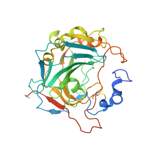
    Tegumental carbonic anhydrase from the worm Schistosoma mansoni (SmCA) is considered a new anti-parasitic target because suppressing its expression interferes with schistosome metabolism and virulence. Here, we present the inhibition profiles of selenoureido compounds on recombinant SmCA and resolution of the first X-ray crystal structures of SmCA in adduct with a selection of such inhibitors. The key molecular features of such compounds in adduct with SmCA were obtained and compared to the human isoform hCA II, in order to understand the main structural factors responsible for enzymatic affinity and selectivity. Compounds that more specifically inhibited the schistosome versus human enzymes were identified. The results expand current knowledge in the field and pave the way for the development of more potent antiparasitic agents in the near future.


  • Organizational Affiliation
    • NEUROFARBA Department, Sezione di Scienze Farmaceutiche, University of Florence, Via Ugo Schiff 6, Sesto Fiorentino, 50019 Florence, Italy.

Macromolecule Content 

  • Total Structure Weight: 73.86 kDa 
  • Atom Count: 5,042 
  • Modeled Residue Count: 557 
  • Deposited Residue Count: 624 
  • Unique protein chains: 1

Macromolecules

Find similar proteins by:|  3D Structure
Entity ID: 1
MoleculeChains  Sequence LengthOrganismDetailsImage
Carbonic anhydrase
A, B
312Schistosoma mansoniMutation(s): 0 
EC: 4.2.1.1
UniProt
Find proteins for A0A3Q0KSG2 (Schistosoma mansoni)
Explore A0A3Q0KSG2 
Go to UniProtKB:  A0A3Q0KSG2
Entity Groups
Sequence Clusters30% Identity50% Identity70% Identity90% Identity95% Identity100% Identity
UniProt GroupA0A3Q0KSG2
Glycosylation
Glycosylation Sites: 3
Sequence Annotations
Expand
Reference Sequence

Oligosaccharides

Help  
Entity ID: 2
MoleculeChains Length2D Diagram GlycosylationD Interactions
2-acetamido-2-deoxy-beta-D-glucopyranose-(1-4)-2-acetamido-2-deoxy-beta-D-glucopyranoseC [auth G]2N/AN-Glycosylation
Glycosylation Resources
GlyTouCan: G42666HT
GlyCosmos: G42666HT
GlyGen: G42666HT

Small Molecules

Ligands 4 Unique
IDChains Name / Formula / InChI Key2D Diagram3D Interactions
TKE
(Subject of Investigation/LOI)

Query on TKE


Download:Ideal Coordinates CCD File
G [auth A],
K [auth B]
1-(4-iodophenyl)-3-[2-(4-sulfamoylphenyl)ethyl]thiourea
C15 H16 I N3 O2 S2
JUSYUNJKOKPYPO-UHFFFAOYSA-N
NAG

Query on NAG


Download:Ideal Coordinates CCD File
D [auth A],
H [auth B],
I [auth B],
L [auth G]
2-acetamido-2-deoxy-beta-D-glucopyranose
C8 H15 N O6
OVRNDRQMDRJTHS-FMDGEEDCSA-N
GOL

Query on GOL


Download:Ideal Coordinates CCD File
F [auth A]GLYCEROL
C3 H8 O3
PEDCQBHIVMGVHV-UHFFFAOYSA-N
ZN

Query on ZN


Download:Ideal Coordinates CCD File
E [auth A],
J [auth B]
ZINC ION
Zn
PTFCDOFLOPIGGS-UHFFFAOYSA-N

Experimental Data & Validation

Experimental Data

  • Method: X-RAY DIFFRACTION
  • Resolution: 2.00 Å
  • R-Value Free:  0.212 (Depositor), 0.215 (DCC) 
  • R-Value Work:  0.168 (Depositor) 
  • R-Value Observed: 0.170 (Depositor) 
Space Group: P 32 2 1
Unit Cell:
Length ( Å )Angle ( ˚ )
a = 103.53α = 90
b = 103.53β = 90
c = 133.11γ = 120
Software Package:
Software NamePurpose
XSCALEdata scaling
REFMACrefinement
PDB_EXTRACTdata extraction
XDSdata reduction
REFMACphasing

Structure Validation

View Full Validation Report


Ligand Structure Quality Assessment 


Entry History 

Deposition Data

Revision History  (Full details and data files)

  • Version 1.0: 2022-04-13
    Type: Initial release
  • Version 1.1: 2022-05-04
    Changes: Database references
  • Version 1.2: 2024-01-31
    Changes: Data collection, Refinement description
  • Version 1.3: 2024-10-23
    Changes: Structure summary