7O45 | pdb_00007o45

Crystal structure of ADD domain of the human DNMT3B methyltransferase


Experimental Data Snapshot

  • Method: X-RAY DIFFRACTION
  • Resolution: 2.10 Å
  • R-Value Free: 
    0.228 (Depositor), 0.231 (DCC) 
  • R-Value Work: 
    0.200 (Depositor), 0.202 (DCC) 
  • R-Value Observed: 
    0.202 (Depositor) 

Starting Model: experimental
View more details

wwPDB Validation 3D Report Full Report

Validation slider image for 7O45

This is version 1.2 of the entry. See complete history

Literature

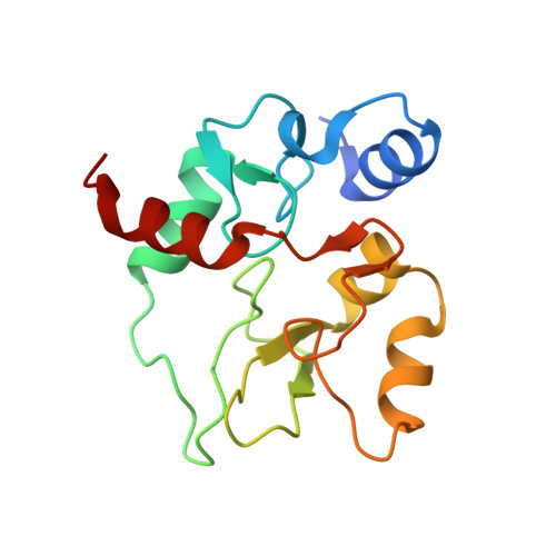
Structure of the DNMT3B ADD domain suggests the absence of a DNMT3A-like autoinhibitory mechanism.

Boyko, K.Arkova, O.Nikolaeva, A.Popov, V.O.Georgiev, P.Bonchuk, A.

(2022) Biochem Biophys Res Commun 619: 124-129

  • DOI: https://doi.org/10.1016/j.bbrc.2022.06.036
  • Primary Citation Related Structures: 
    7O45

  • PubMed Abstract: 

    De novo DNA methylation in early mammalian development depends on the activity of the DNMT3 methyltransferase family. An autoinhibitory mechanism involving the interaction between ADD and the catalytic domains of DNMT3A has been described. ADD is a zinc-coordinating histone-binding domain. The ADD domain of DNMT3A, when bound to a K4-unmethylated histone H3 tail, switches the enzyme to its catalytically active state. DNMT3B is another de novo methyltransferase enzyme with a more strict tissue- and stage-specific expression profile and a slightly different site specificity, lacking cooperative DNA methylation activity. Here, we obtained the crystal structure of the DNMT3B ADD domain, which demonstrated the extended conformation of the autoinhibitory loop even in the absence of the histone H3 tail. The lack of interaction between DNMT3B ADD and the methyltransferase domain was confirmed using an in vitro pull-down assay. The structural rearrangements in the loop also created an additional protein interaction interface leading to the formation of trimers in crystal, which may reflect their possible involvement in some unknown protein-protein interactions. Our results suggest that DNMT3B, in contrast to DNMT3A, has different modes of regulation of its activity that are independent of H3K4 methylation status.


  • Organizational Affiliation
    • Department of the Control of Genetic Processes, Institute of Gene Biology Russian Academy of Sciences, 34/5 Vavilov St., Moscow, 119334, Russia; Bach Institute of Biochemistry, Research Center of Biotechnology Russian Academy of Sciences, Leninsky pr-t, 33, Bld. 2, Moscow, 119071, Russia.

Macromolecule Content 

  • Total Structure Weight: 67.25 kDa 
  • Atom Count: 4,065 
  • Modeled Residue Count: 507 
  • Deposited Residue Count: 584 
  • Unique protein chains: 1

Macromolecules

Find similar proteins by:|  3D Structure
Entity ID: 1
MoleculeChains  Sequence LengthOrganismDetailsImage
Isoform 6 of DNA (cytosine-5)-methyltransferase 3B
A, B, C, D
146Homo sapiensMutation(s): 0 
Gene Names: DNMT3B
EC: 2.1.1.37
UniProt & NIH Common Fund Data Resources
Find proteins for Q9UBC3 (Homo sapiens)
Explore Q9UBC3 
Go to UniProtKB:  Q9UBC3
PHAROS:  Q9UBC3
GTEx:  ENSG00000088305 
Entity Groups
Sequence Clusters30% Identity50% Identity70% Identity90% Identity95% Identity100% Identity
UniProt GroupQ9UBC3
Sequence Annotations
Expand
Reference Sequence

Small Molecules

Ligands 2 Unique
IDChains Name / Formula / InChI Key2D Diagram3D Interactions
BR

Query on BR


Download:Ideal Coordinates CCD File
H [auth A],
I [auth A],
P [auth C]
BROMIDE ION
Br
CPELXLSAUQHCOX-UHFFFAOYSA-M
ZN
(Subject of Investigation/LOI)

Query on ZN


Download:Ideal Coordinates CCD File
E [auth A]
F [auth A]
G [auth A]
J [auth B]
K [auth B]
E [auth A],
F [auth A],
G [auth A],
J [auth B],
K [auth B],
L [auth B],
M [auth C],
N [auth C],
O [auth C],
Q [auth D],
R [auth D],
S [auth D]
ZINC ION
Zn
PTFCDOFLOPIGGS-UHFFFAOYSA-N

Experimental Data & Validation

Experimental Data

  • Method: X-RAY DIFFRACTION
  • Resolution: 2.10 Å
  • R-Value Free:  0.228 (Depositor), 0.231 (DCC) 
  • R-Value Work:  0.200 (Depositor), 0.202 (DCC) 
  • R-Value Observed: 0.202 (Depositor) 
Space Group: P 21 21 21
Unit Cell:
Length ( Å )Angle ( ˚ )
a = 80.282α = 90
b = 89.943β = 90
c = 92.195γ = 90
Software Package:
Software NamePurpose
Aimlessdata scaling
PHASERphasing
REFMACrefinement
PDB_EXTRACTdata extraction
DIALSdata reduction

Structure Validation

View Full Validation Report


Entry History 

& Funding Information

Deposition Data


Funding OrganizationLocationGrant Number
Russian Science FoundationRussian Federation19-74-10099

Revision History  (Full details and data files)

  • Version 1.0: 2022-04-13
    Type: Initial release
  • Version 1.1: 2022-10-26
    Changes: Database references
  • Version 1.2: 2024-01-31
    Changes: Data collection, Refinement description