7O30

Crystal structure of the anti-PAS Fab 1.1 in complex with its epitope peptide


Experimental Data Snapshot

  • Method: X-RAY DIFFRACTION
  • Resolution: 2.65 Å
  • R-Value Free: 0.269 
  • R-Value Work: 0.220 
  • R-Value Observed: 0.222 

wwPDB Validation   3D Report Full Report


This is version 1.2 of the entry. See complete history


Literature

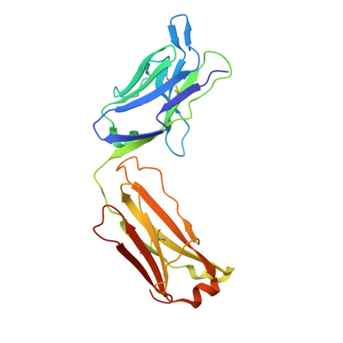
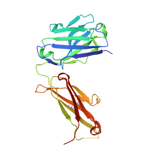
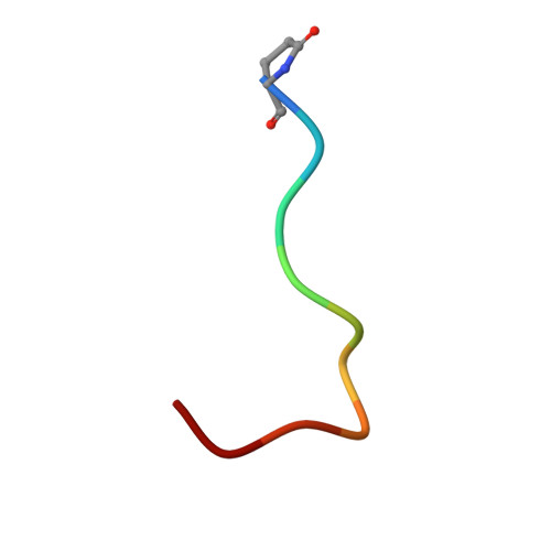
Molecular recognition of structurally disordered Pro/Ala-rich sequences (PAS) by antibodies involves an Ala residue at the hot spot of the epitope.

Schilz, J.Binder, U.Friedrich, L.Gebauer, M.Lutz, C.Schlapschy, M.Schiefner, A.Skerra, A.

(2021) J Mol Biol 433: 167113-167113

  • DOI: https://doi.org/10.1016/j.jmb.2021.167113
  • Primary Citation of Related Structures:  
    7O2Z, 7O30, 7O31, 7O33

  • PubMed Abstract: 

    Pro/Ala-rich sequences (PAS) are polypeptides that were developed as a biological alternative to poly-ethylene glycol (PEG) to generate biopharmaceuticals with extended plasma half-life. Like PEG, PAS polypeptides are conformationally disordered and show high solubility in water. Devoid of any charged or prominent hydrophobic side chains, these biosynthetic polymers represent an extreme case of intrinsically disordered proteins. Despite lack of immunogenicity of PAS tags in numerous animal studies we now succeeded in generating monoclonal antibodies (MAbs) against three different PAS versions. To this end, mice were immunized with a PAS#1, P/A#1 or APSA 40mer peptide conjugated to keyhole limpet hemocyanin as highly immunogenic carrier protein. In each case, one MAb with high binding activity and specificity towards a particular PAS motif was obtained. The apparent affinity was strongly dependent on the avidity effect and most pronounced for the bivalent MAb when interacting with a long PAS repeat. X-ray structural analysis of four representative anti-PAS Fab fragments in complex with their cognate PAS epitope peptides revealed interactions dominated by hydrogen bond networks involving the peptide backbone as well as multiple Van der Waals contacts arising from intimate shape complementarity. Surprisingly, Ala, the L-amino acid with the smallest side chain, emerged as a crucial feature for epitope recognition, contributing specific contacts at the center of the paratope in several anti-PAS complexes. Apart from these insights into how antibodies can recognize feature-less peptides without secondary structure, the MAbs characterized in this study offer valuable reagents for the preclinical and clinical development of PASylated biologics.


  • Organizational Affiliation

    Lehrstuhl für Biologische Chemie, Technische Universität München, Emil-Erlenmeyer-Forum 5, 85354 Freising, Germany.


Macromolecules
Find similar proteins by:  (by identity cutoff)  |  3D Structure
Entity ID: 1
MoleculeChains Sequence LengthOrganismDetailsImage
anti-PAS Fab 1.1 chimeric light chainA [auth L],
D [auth A]
218Mus musculusMutation(s): 0 
Entity Groups  
Sequence Clusters30% Identity50% Identity70% Identity90% Identity95% Identity100% Identity
Sequence Annotations
Expand
  • Reference Sequence
Find similar proteins by:  (by identity cutoff)  |  3D Structure
Entity ID: 2
MoleculeChains Sequence LengthOrganismDetailsImage
anti-PAS Fab 1.1 chimeric heavy chainB [auth H],
E [auth B]
229Mus musculusMutation(s): 0 
Entity Groups  
Sequence Clusters30% Identity50% Identity70% Identity90% Identity95% Identity100% Identity
Sequence Annotations
Expand
  • Reference Sequence

Find similar proteins by:  Sequence   |   3D Structure  

Entity ID: 3
MoleculeChains Sequence LengthOrganismDetailsImage
PAS#1 epitope peptideC [auth P],
F [auth Q]
10synthetic constructMutation(s): 0 
Entity Groups  
Sequence Clusters30% Identity50% Identity70% Identity90% Identity95% Identity100% Identity
Sequence Annotations
Expand
  • Reference Sequence
Small Molecules
Modified Residues  1 Unique
IDChains TypeFormula2D DiagramParent
PCA
Query on PCA
C [auth P],
F [auth Q]
L-PEPTIDE LINKINGC5 H7 N O3GLN
Experimental Data & Validation

Experimental Data

  • Method: X-RAY DIFFRACTION
  • Resolution: 2.65 Å
  • R-Value Free: 0.269 
  • R-Value Work: 0.220 
  • R-Value Observed: 0.222 
  • Space Group: P 42 21 2
Unit Cell:
Length ( Å )Angle ( ˚ )
a = 102.615α = 90
b = 102.615β = 90
c = 199.85γ = 90
Software Package:
Software NamePurpose
XDSdata reduction
XSCALEdata scaling
PHASERphasing
REFMACrefinement
PDB_EXTRACTdata extraction

Structure Validation

View Full Validation Report


Entry History 

Deposition Data

Revision History  (Full details and data files)

  • Version 1.0: 2021-07-07
    Type: Initial release
  • Version 1.1: 2021-07-21
    Changes: Database references
  • Version 1.2: 2024-01-31
    Changes: Data collection, Database references, Refinement description