7NXG | pdb_00007nxg

Wax synthase 1 from Acinetobacter baylyi (AbWSD1) co-crystallized with myristic acid


Experimental Data Snapshot

  • Method: X-RAY DIFFRACTION
  • Resolution: 1.95 Å
  • R-Value Free: 
    0.209 (Depositor), 0.209 (DCC) 
  • R-Value Work: 
    0.189 (Depositor), 0.190 (DCC) 
  • R-Value Observed: 
    0.190 (Depositor) 

wwPDB Validation 3D Report Full Report

Validation slider image for 7NXG

Ligand Structure Quality Assessment 


This is version 2.1 of the entry. See complete history

Literature

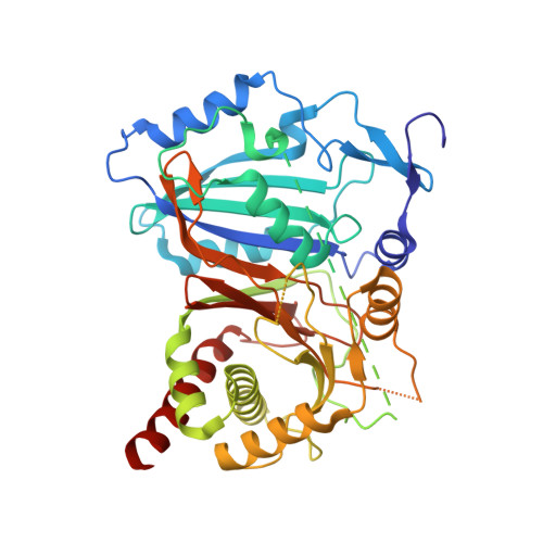
Crystal Structure of the Bifunctional Wax Synthase 1 from Acinetobacter baylyi Suggests a Conformational Change upon Substrate Binding and Formation of Additional Substrate Binding Sites

Vollheyde, K.Kuhnel, K.Lambrecht, F.Kawelke, S.Herrfurth, C.Feussner, I.

(2022) ACS Catal 12: 9753-9765

Macromolecule Content 

  • Total Structure Weight: 52.59 kDa 
  • Atom Count: 3,312 
  • Modeled Residue Count: 390 
  • Deposited Residue Count: 463 
  • Unique protein chains: 1

Macromolecules

Find similar proteins by:|  3D Structure
Entity ID: 1
MoleculeChains  Sequence LengthOrganismDetailsImage
O-acyltransferase WSD463Acinetobacter baylyi ADP1Mutation(s): 0 
Gene Names: wax-dgaTACIAD0832
EC: 2.3.1.20 (PDB Primary Data), 2.3.1.75 (PDB Primary Data)
UniProt
Find proteins for Q8GGG1 (Acinetobacter baylyi (strain ATCC 33305 / BD413 / ADP1))
Explore Q8GGG1 
Go to UniProtKB:  Q8GGG1
Entity Groups
Sequence Clusters30% Identity50% Identity70% Identity90% Identity95% Identity100% Identity
UniProt GroupQ8GGG1
Sequence Annotations
Expand
Reference Sequence

Small Molecules

Ligands 1 Unique
IDChains Name / Formula / InChI Key2D Diagram3D Interactions
MYR
(Subject of Investigation/LOI)

Query on MYR


Download:Ideal Coordinates CCD File
B [auth A]MYRISTIC ACID
C14 H28 O2
TUNFSRHWOTWDNC-UHFFFAOYSA-N

Experimental Data & Validation

Experimental Data

  • Method: X-RAY DIFFRACTION
  • Resolution: 1.95 Å
  • R-Value Free:  0.209 (Depositor), 0.209 (DCC) 
  • R-Value Work:  0.189 (Depositor), 0.190 (DCC) 
  • R-Value Observed: 0.190 (Depositor) 
Space Group: I 4 2 2
Unit Cell:
Length ( Å )Angle ( ˚ )
a = 118.32α = 90
b = 118.32β = 90
c = 142.56γ = 90
Software Package:
Software NamePurpose
PHENIXrefinement
XDSdata reduction
XSCALEdata scaling
PDB_EXTRACTdata extraction
SHELXCDphasing

Structure Validation

View Full Validation Report


Ligand Structure Quality Assessment 


Entry History 

& Funding Information

Deposition Data


Funding OrganizationLocationGrant Number
German Research Foundation (DFG)Germany--

Revision History  (Full details and data files)

  • Version 1.0: 2022-03-30
    Type: Initial release
  • Version 2.0: 2022-10-12
    Changes: Atomic model, Database references, Source and taxonomy
  • Version 2.1: 2024-06-19
    Changes: Data collection