


The cryo-EM structure of the bacterial type I segregation filament reveals ParA s conformational plasticity upon DNA binding
Parker, A.V., Mann, D., Tzokov, S.B., Hwang, L.C., Bergeron, J.R.C.To be published.
Experimental Data Snapshot
Starting Model: experimental
View more details
Entity ID: 1 | |||||
|---|---|---|---|---|---|
| Molecule | Chains | Sequence Length | Organism | Details | Image |
| AAA family ATPase | 407 | Vibrio cholerae | Mutation(s): 0 Gene Names: parA, BC353_10845, C9J66_03480, ERS013138_01197, ERS013166_00021, ERS013186_00500, ERS013193_00027, ERS013197_04093, ERS013198_00323, ERS013199_01186... | 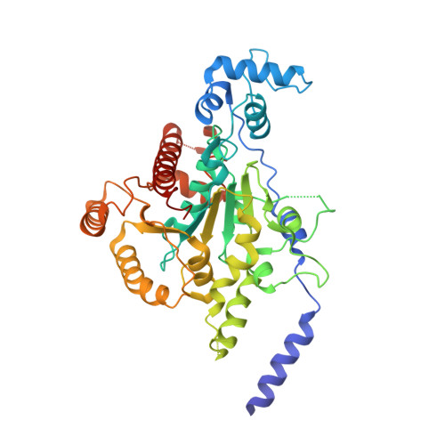 | |
UniProt | |||||
Entity Groups | |||||
| Sequence Clusters | 30% Identity50% Identity70% Identity90% Identity95% Identity100% Identity | ||||
| UniProt Group | Q9KKJ2 | ||||
Sequence AnnotationsExpand | |||||
Reference Sequence | |||||
| Ligands 3 Unique | |||||
|---|---|---|---|---|---|
| ID | Chains | Name / Formula / InChI Key | 2D Diagram | 3D Interactions | |
| ADP (Subject of Investigation/LOI) Download:Ideal Coordinates CCD File | E [auth A], H [auth B], K [auth C], M [auth D] | ADENOSINE-5'-DIPHOSPHATE C10 H15 N5 O10 P2 XTWYTFMLZFPYCI-KQYNXXCUSA-N |  | ||
| GOL Download:Ideal Coordinates CCD File | G [auth A], J [auth B] | GLYCEROL C3 H8 O3 PEDCQBHIVMGVHV-UHFFFAOYSA-N |  | ||
| MG Download:Ideal Coordinates CCD File | F [auth A], I [auth B], L [auth C], N [auth D] | MAGNESIUM ION Mg JLVVSXFLKOJNIY-UHFFFAOYSA-N |  | ||
| Length ( Å ) | Angle ( ˚ ) |
|---|---|
| a = 199.205 | α = 90 |
| b = 199.205 | β = 90 |
| c = 260.066 | γ = 120 |
| Software Name | Purpose |
|---|---|
| PHENIX | refinement |
| autoPROC | data scaling |
| PHASER | phasing |
| PDB_EXTRACT | data extraction |
| autoPROC | data reduction |
| Funding Organization | Location | Grant Number |
|---|---|---|
| Not funded | -- |


