The role of streptavidin and its variants in catalysis by biotinylated secondary amines.
Nodling, A.R., Santi, N., Castillo, R., Lipka-Lloyd, M., Jin, Y., Morrill, L.C., Swiderek, K., Moliner, V., Luk, L.Y.P.(2021) Org Biomol Chem 19: 10424-10431
- PubMed: 34825690 Search on PubMedSearch on PubMed Central
- DOI: https://doi.org/10.1039/d1ob01947c
- Primary Citation Related Structures:
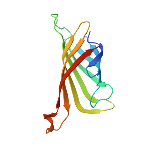
6ZYT, 7NLV - PubMed Abstract:
Here, we combine the use of host screening, protein crystallography and QM/MM molecular dynamics simulations to investigate how the protein structure affects iminium catalysis by biotinylated secondary amines in a model 1,4 conjugate addition reaction. Monomeric streptavidin (M-Sav) lacks a quaternary structure and the solvent-exposed reaction site resulted in poor product conversion in the model reaction with low enantio- and regioselectivities. These parameters were much improved when the tetrameric host T-Sav was used; indeed, residues at the symmetrical subunit interface were proven to be critical for catalysis through a mutagenesis study. The use of QM/MM simulations and the asymmetric dimeric variant D-Sav revealed that both Lys121 residues which are located in the hosting and neighboring subunits play a critical role in controlling the stereoselectivity and reactivity. Lastly, the D-Sav template, though providing a lower conversion than that of the symmetric tetrameric counterpart, is likely a better starting point for future protein engineering because each surrounding residue within the asymmetric scaffold can be refined for secondary amine catalysis.
- School of Chemistry, Main Building, Cardiff University, Cardiff, CF10 3AT, UK. LukLY@cardiff.ac.uk.
Organizational Affiliation: