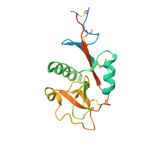
Sweet Drugs for Bad Bugs: A Glycomimetic Strategy against the DC-SIGN-Mediated Dissemination of SARS-CoV-2.
Cramer, J., Lakkaichi, A., Aliu, B., Jakob, R.P., Klein, S., Cattaneo, I., Jiang, X., Rabbani, S., Schwardt, O., Zimmer, G., Ciancaglini, M., Abreu Mota, T., Maier, T., Ernst, B.(2021) J Am Chem Soc 143: 17465-17478
- PubMed: 34652144
- DOI: https://doi.org/10.1021/jacs.1c06778
- Primary Citation of Related Structures:
7NL6, 7NL7 - PubMed Abstract:
The C-type lectin receptor DC-SIGN is a pattern recognition receptor expressed on macrophages and dendritic cells. It has been identified as a promiscuous entry receptor for many pathogens, including epidemic and pandemic viruses such as SARS-CoV-2, Ebola virus, and HIV-1. In the context of the recent SARS-CoV-2 pandemic, DC-SIGN-mediated virus dissemination and stimulation of innate immune responses has been implicated as a potential factor in the development of severe COVID-19. Inhibition of virus binding to DC-SIGN, thus, represents an attractive host-directed strategy to attenuate overshooting innate immune responses and prevent the progression of the disease. In this study, we report on the discovery of a new class of potent glycomimetic DC-SIGN antagonists from a focused library of triazole-based mannose analogues. Structure-based optimization of an initial screening hit yielded a glycomimetic ligand with a more than 100-fold improved binding affinity compared to methyl α-d-mannopyranoside. Analysis of binding thermodynamics revealed an enthalpy-driven improvement of binding affinity that was enabled by hydrophobic interactions with a loop region adjacent to the binding site and displacement of a conserved water molecule. The identified ligand was employed for the synthesis of multivalent glycopolymers that were able to inhibit SARS-CoV-2 spike glycoprotein binding to DC-SIGN-expressing cells, as well as DC-SIGN-mediated trans -infection of ACE2 + cells by SARS-CoV-2 spike protein-expressing viruses, in nanomolar concentrations. The identified glycomimetic ligands reported here open promising perspectives for the development of highly potent and fully selective DC-SIGN-targeted therapeutics for a broad spectrum of viral infections.
- University of Basel, Institute of Molecular Pharmacy, Pharmacenter of the University of Basel, Klingelbergstrasse 50, 4056 Basel, Switzerland.
Organizational Affiliation:


