7NCX | pdb_00007ncx

Crystal structure of GH30 (double mutant EE) from Thermothelomyces thermophila.


Experimental Data Snapshot

  • Method: X-RAY DIFFRACTION
  • Resolution: 1.36 Å
  • R-Value Free: 
    0.158 (Depositor), 0.144 (DCC) 
  • R-Value Work: 
    0.122 (Depositor) 

Starting Model: experimental
View more details

wwPDB Validation 3D Report Full Report

Validation slider image for 7NCX

Ligand Structure Quality Assessment 


This is version 1.2 of the entry. See complete history

Literature

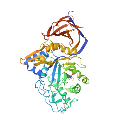
Unique features of the bifunctional GH30 from Thermothelomyces thermophila revealed by structural and mutational studies

Nikolaivits, E.Pentari, C.Kosinas, C.Feiler, C.G.Spiliopoulou, M.Weiss, M.S.Dimarogona, M.Topakas, E.

(2021) Carbohydrate Polymers 273: 118553

Macromolecule Content 

  • Total Structure Weight: 53.35 kDa 
  • Atom Count: 4,082 
  • Modeled Residue Count: 443 
  • Deposited Residue Count: 482 
  • Unique protein chains: 1

Macromolecules

Find similar proteins by:|  3D Structure
Entity ID: 1
MoleculeChains  Sequence LengthOrganismDetailsImage
GH30 family xylanaseA [auth AAA]482Thermothelomyces thermophilus ATCC 42464Mutation(s): 2 
Gene Names: Xyn30AMYCTH_38558
EC: 3.2.1
UniProt
Find proteins for G2Q1N4 (Thermothelomyces thermophilus (strain ATCC 42464 / BCRC 31852 / DSM 1799))
Explore G2Q1N4 
Go to UniProtKB:  G2Q1N4
Entity Groups
Sequence Clusters30% Identity50% Identity70% Identity90% Identity95% Identity100% Identity
UniProt GroupG2Q1N4
Glycosylation
Glycosylation Sites: 2
Sequence Annotations
Expand
Reference Sequence

Oligosaccharides

Help  
Entity ID: 2
MoleculeChains Length2D Diagram GlycosylationD Interactions
alpha-D-mannopyranose-(1-3)-alpha-D-mannopyranose-(1-6)-[alpha-D-mannopyranose-(1-3)]beta-D-mannopyranose-(1-4)-2-acetamido-2-deoxy-beta-D-glucopyranose-(1-4)-2-acetamido-2-deoxy-beta-D-glucopyranoseB [auth BBB]6N-Glycosylation
Glycosylation Resources
GlyTouCan: G09724ZC
GlyCosmos: G09724ZC
GlyGen: G09724ZC

Small Molecules

Ligands 4 Unique
IDChains Name / Formula / InChI Key2D Diagram3D Interactions
NAG

Query on NAG


Download:Ideal Coordinates CCD File
G [auth AAA]2-acetamido-2-deoxy-beta-D-glucopyranose
C8 H15 N O6
OVRNDRQMDRJTHS-FMDGEEDCSA-N
PEG

Query on PEG


Download:Ideal Coordinates CCD File
D [auth AAA]DI(HYDROXYETHYL)ETHER
C4 H10 O3
MTHSVFCYNBDYFN-UHFFFAOYSA-N
GOL

Query on GOL


Download:Ideal Coordinates CCD File
C [auth AAA]GLYCEROL
C3 H8 O3
PEDCQBHIVMGVHV-UHFFFAOYSA-N
EDO

Query on EDO


Download:Ideal Coordinates CCD File
E [auth AAA],
F [auth AAA]
1,2-ETHANEDIOL
C2 H6 O2
LYCAIKOWRPUZTN-UHFFFAOYSA-N

Experimental Data & Validation

Experimental Data

  • Method: X-RAY DIFFRACTION
  • Resolution: 1.36 Å
  • R-Value Free:  0.158 (Depositor), 0.144 (DCC) 
  • R-Value Work:  0.122 (Depositor) 
Space Group: P 21 21 21
Unit Cell:
Length ( Å )Angle ( ˚ )
a = 38.887α = 90
b = 87.315β = 90
c = 107.917γ = 90
Software Package:
Software NamePurpose
REFMACrefinement
Aimlessdata reduction
XDSdata scaling
PHASERphasing

Structure Validation

View Full Validation Report


Ligand Structure Quality Assessment 


Entry History 

Deposition Data

Revision History  (Full details and data files)

  • Version 1.0: 2021-09-08
    Type: Initial release
  • Version 1.1: 2024-01-31
    Changes: Data collection, Derived calculations, Refinement description
  • Version 1.2: 2024-11-13
    Changes: Structure summary