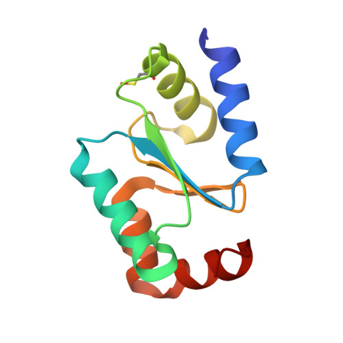
Atypical Iron-Sulfur Cluster Binding, Redox Activity and Structural Properties of Chlamydomonas reinhardtii Glutaredoxin 2.
Roret, T., Zhang, B., Moseler, A., Dhalleine, T., Gao, X.H., Couturier, J., Lemaire, S.D., Didierjean, C., Johnson, M.K., Rouhier, N.(2021) Antioxidants (Basel) 10
- PubMed: 34069657
- DOI: https://doi.org/10.3390/antiox10050803
- Primary Citation of Related Structures:
7NCV, 7NCW - PubMed Abstract:
Glutaredoxins (GRXs) are thioredoxin superfamily members exhibiting thiol-disulfide oxidoreductase activity and/or iron-sulfur (Fe-S) cluster binding capacities. These properties are determined by specific structural factors. In this study, we examined the capacity of the class I Chlamydomonas reinhardtii GRX2 recombinant protein to catalyze both protein glutathionylation and deglutathionylation reactions using a redox sensitive fluorescent protein as a model protein substrate. We observed that the catalytic cysteine of the CPYC active site motif of GRX2 was sufficient for catalyzing both reactions in the presence of glutathione. Unexpectedly, spectroscopic characterization of the protein purified under anaerobiosis showed the presence of a [2Fe-2S] cluster despite having a presumably inadequate active site signature, based on past mutational analyses. The spectroscopic characterization of cysteine mutated variants together with modeling of the Fe-S cluster-bound GRX homodimer from the structure of an apo-GRX2 indicate the existence of an atypical Fe-S cluster environment and ligation mode. Overall, the results further delineate the biochemical and structural properties of conventional GRXs, pointing to the existence of multiple factors more complex than anticipated, sustaining the capacity of these proteins to bind Fe-S clusters.
- Université de Lorraine, INRAE, IAM, F-54000 Nancy, France.
Organizational Affiliation:


