7N3R

The ternary complex of human Bisphosphoglycerate mutase with 3-phosphoglycerate and 2-phosphoglycolate


Experimental Data Snapshot

  • Method: X-RAY DIFFRACTION
  • Resolution: 2.25 Å
  • R-Value Free: 0.274 
  • R-Value Work: 0.203 
  • R-Value Observed: 0.206 

wwPDB Validation   3D Report Full Report


Ligand Structure Quality Assessment 


This is version 1.1 of the entry. See complete history


Literature

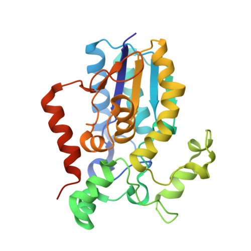
Molecular insight into 2-phosphoglycolate activation of the phosphatase activity of bisphosphoglycerate mutase.

Aljahdali, A.S.Musayev, F.N.Burgner 2nd, J.W.Ghatge, M.S.Shekar, V.Zhang, Y.Omar, A.M.Safo, M.K.

(2022) Acta Crystallogr D Struct Biol 78: 472-482

  • DOI: https://doi.org/10.1107/S2059798322001802
  • Primary Citation of Related Structures:  
    7N3R, 7N3S

  • PubMed Abstract: 

    Bisphosphoglycerate mutase (BPGM) is an erythrocyte-specific multifunctional enzyme that is responsible for the regulation of 2,3-bisphosphoglycerate (2,3-BPG) in red blood cells through its synthase and phosphatase activities; the latter enzymatic function is stimulated by the endogenous activator 2-phosphoglycolate (2-PG). 2,3-BPG is a natural allosteric effector of hemoglobin (Hb) that is responsible for decreasing the affinity of Hb for oxygen to facilitate tissue oxygenation. Here, crystal structures of BPGM with 2-PG in the presence and absence of 3-phosphoglycerate are reported at 2.25 and 2.48 Å resolution, respectively. Structure analysis revealed a new binding site for 2-PG at the dimer interface for the first time, in addition to the expected active-site binding. Also, conformational non-equivalence of the two active sites was observed as one of the sites was found in an open conformation, with the residues at the active-site entrance, including Arg100, Arg116 and Arg117, and the C-terminus disordered. The kinetic result is consistent with the binding of 2-PG to an allosteric or noncatalytic site as well as the active site. This study paves the way for the rational targeting of BPGM for therapeutic purposes, especially for the treatment of sickle cell disease.


  • Organizational Affiliation

    Department of Pharmaceutical Chemistry, King Abdulaziz University, Alsulaymanyah, Jeddah 21589, Saudi Arabia.


Macromolecules
Find similar proteins by:  (by identity cutoff)  |  3D Structure
Entity ID: 1
MoleculeChains Sequence LengthOrganismDetailsImage
Bisphosphoglycerate mutase
A, B
267Homo sapiensMutation(s): 0 
Gene Names: BPGM
EC: 5.4.2.4 (PDB Primary Data), 5.4.2.11 (PDB Primary Data)
UniProt & NIH Common Fund Data Resources
Find proteins for P07738 (Homo sapiens)
Explore P07738 
Go to UniProtKB:  P07738
PHAROS:  P07738
GTEx:  ENSG00000172331 
Entity Groups  
Sequence Clusters30% Identity50% Identity70% Identity90% Identity95% Identity100% Identity
UniProt GroupP07738
Sequence Annotations
Expand
  • Reference Sequence
Experimental Data & Validation

Experimental Data

  • Method: X-RAY DIFFRACTION
  • Resolution: 2.25 Å
  • R-Value Free: 0.274 
  • R-Value Work: 0.203 
  • R-Value Observed: 0.206 
  • Space Group: P 21 21 21
Unit Cell:
Length ( Å )Angle ( ˚ )
a = 53.072α = 90
b = 70.865β = 90
c = 159.803γ = 90
Software Package:
Software NamePurpose
Aimlessdata scaling
PHASERphasing
PHENIXrefinement
PDB_EXTRACTdata extraction
CrysalisProdata reduction

Structure Validation

View Full Validation Report


Ligand Structure Quality Assessment 


Entry History & Funding Information

Deposition Data


Funding OrganizationLocationGrant Number
National Institutes of Health/National Institute on Minority Health and Health Disparities (NIH/NIMHD)United StatesR01MD009124

Revision History  (Full details and data files)

  • Version 1.0: 2022-04-13
    Type: Initial release
  • Version 1.1: 2023-10-18
    Changes: Data collection, Refinement description