7MYL

Crystal structure of DfrA1 dihydrofolate reductase in complex with TRIMETHOPRIM


Experimental Data Snapshot

  • Method: X-RAY DIFFRACTION
  • Resolution: 2.15 Å
  • R-Value Free: 0.266 
  • R-Value Work: 0.215 
  • R-Value Observed: 0.217 

wwPDB Validation   3D Report Full Report


Ligand Structure Quality Assessment 


This is version 1.2 of the entry. See complete history


Literature

Structure-guided functional studies of plasmid-encoded dihydrofolate reductases reveal a common mechanism of trimethoprim resistance in Gram-negative pathogens.

Krucinska, J.Lombardo, M.N.Erlandsen, H.Estrada, A.Si, D.Viswanathan, K.Wright, D.L.

(2022) Commun Biol 5: 459-459

  • DOI: https://doi.org/10.1038/s42003-022-03384-y
  • Primary Citation of Related Structures:  
    7MQP, 7MYL, 7MYM, 7NAE, 7R6G, 7REB, 7REG, 7RGJ, 7RGK, 7RGO

  • PubMed Abstract: 

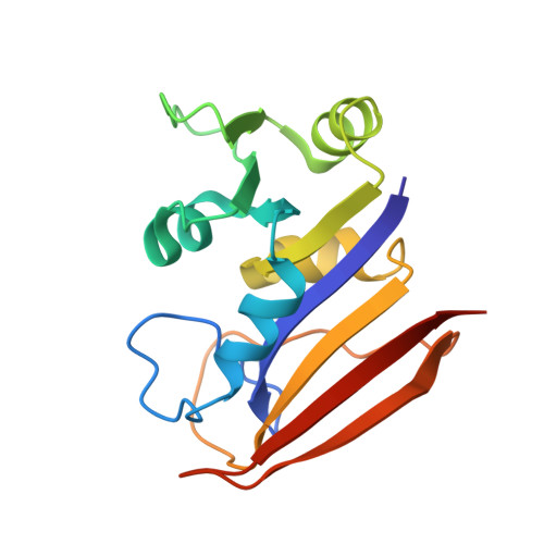
    Two plasmid-encoded dihydrofolate reductase (DHFR) isoforms, DfrA1 and DfrA5, that give rise to high levels of resistance in Gram-negative bacteria were structurally and biochemically characterized to reveal the mechanism of TMP resistance and to support phylogenic groupings for drug development against antibiotic resistant pathogens. Preliminary screening of novel antifolates revealed related chemotypes that showed high levels of inhibitory potency against Escherichia coli chromosomal DHFR (EcDHFR), DfrA1, and DfrA5. Kinetics and biophysical analysis, coupled with crystal structures of trimethoprim bound to EcDHFR, DfrA1 and DfrA5, and two propargyl-linked antifolates (PLA) complexed with EcDHFR, DfrA1 and DfrA5, were determined to define structural features of the substrate binding pocket and guide synthesis of pan-DHFR inhibitors.


  • Organizational Affiliation

    Department of Pharmaceutical Sciences, University of Connecticut, 69N. Eagleville Rd., Storrs, CT, 06269, USA.


Macromolecules
Find similar proteins by:  (by identity cutoff)  |  3D Structure
Entity ID: 1
MoleculeChains Sequence LengthOrganismDetailsImage
Dihydrofolate reductase
A, B, C, D, E
A, B, C, D, E, F
161Klebsiella pneumoniaeMutation(s): 0 
Gene Names: dfrA1AM447_11900EXT45_00270
EC: 1.5.1.3
UniProt
Find proteins for A4GRC7 (Klebsiella pneumoniae)
Explore A4GRC7 
Go to UniProtKB:  A4GRC7
Entity Groups  
Sequence Clusters30% Identity50% Identity70% Identity90% Identity95% Identity100% Identity
UniProt GroupA4GRC7
Sequence Annotations
Expand
  • Reference Sequence
Experimental Data & Validation

Experimental Data

  • Method: X-RAY DIFFRACTION
  • Resolution: 2.15 Å
  • R-Value Free: 0.266 
  • R-Value Work: 0.215 
  • R-Value Observed: 0.217 
  • Space Group: P 1 21 1
Unit Cell:
Length ( Å )Angle ( ˚ )
a = 55.072α = 90
b = 72.89β = 90.56
c = 125.122γ = 90
Software Package:
Software NamePurpose
REFMACrefinement
MOSFLMdata reduction
Aimlessdata scaling
PHASERphasing
PDB_EXTRACTdata extraction

Structure Validation

View Full Validation Report


Ligand Structure Quality Assessment 


Entry History & Funding Information

Deposition Data


Funding OrganizationLocationGrant Number
National Institutes of Health/National Institute Of Allergy and Infectious Diseases (NIH/NIAID)United States1 R01AI111957

Revision History  (Full details and data files)

  • Version 1.0: 2022-06-01
    Type: Initial release
  • Version 1.1: 2022-08-24
    Changes: Database references
  • Version 1.2: 2023-10-18
    Changes: Data collection, Refinement description