7MYH

Ubiquitin variant UbV.k.2 in complex with Ube2k


Experimental Data Snapshot

  • Method: X-RAY DIFFRACTION
  • Resolution: 2.39 Å
  • R-Value Free: 0.249 
  • R-Value Work: 0.206 
  • R-Value Observed: 0.208 

wwPDB Validation   3D Report Full Report


This is version 1.2 of the entry. See complete history


Literature

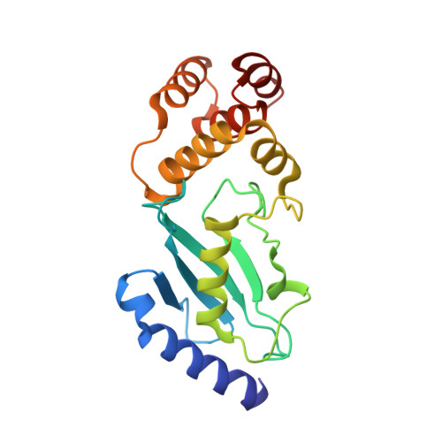
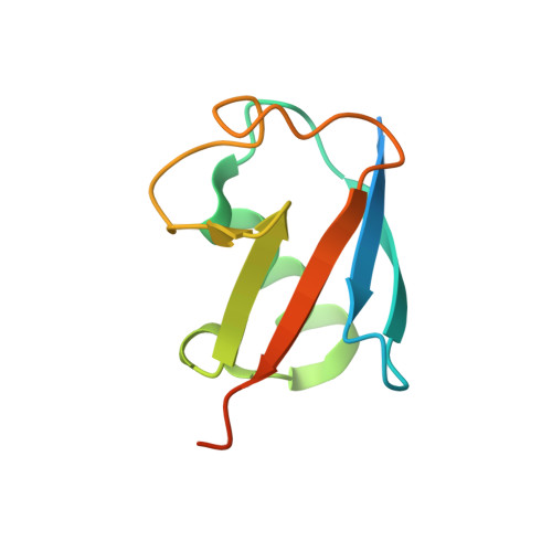
Identification of Ubiquitin Variants That Inhibit the E2 Ubiquitin Conjugating Enzyme, Ube2k.

Middleton, A.J.Teyra, J.Zhu, J.Sidhu, S.S.Day, C.L.

(2021) ACS Chem Biol 16: 1745-1756

  • DOI: https://doi.org/10.1021/acschembio.1c00445
  • Primary Citation of Related Structures:  
    7MYF, 7MYH

  • PubMed Abstract: 

    Transfer of ubiquitin to substrate proteins regulates most processes in eukaryotic cells. E2 enzymes are a central component of the ubiquitin machinery, and generally determine the type of ubiquitin signal generated and thus the ultimate fate of substrate proteins. The E2, Ube2k, specifically builds degradative ubiquitin chains on diverse substrates. Here we have identified protein-based reagents, called ubiquitin variants (UbVs), that bind tightly and specifically to Ube2k. Crystal structures reveal that the UbVs bind to the E2 enzyme at a hydrophobic cleft that is distinct from the active site and previously identified ubiquitin binding sites. We demonstrate that the UbVs are potent inhibitors of Ube2k and block both ubiquitin charging of the E2 enzyme and E3-catalyzed ubiquitin transfer. The binding site of the UbVs suggests they directly clash with the ubiquitin activating enzyme, while potentially disrupting interactions with E3 ligases via allosteric effects. Our data reveal the first protein-based inhibitors of Ube2k and unveil a hydrophobic groove that could be an effective target for inhibiting Ube2k and other E2 enzymes.


  • Organizational Affiliation

    Department of Biochemistry, School of Biomedical Sciences, University of Otago, Dunedin 9054, New Zealand.


Macromolecules
Find similar proteins by:  (by identity cutoff)  |  3D Structure
Entity ID: 1
MoleculeChains Sequence LengthOrganismDetailsImage
Ubiquitin-conjugating enzyme E2 K201Homo sapiensMutation(s): 0 
Gene Names: UBE2KHIP2LIG
EC: 2.3.2.23
UniProt & NIH Common Fund Data Resources
Find proteins for P61086 (Homo sapiens)
Explore P61086 
Go to UniProtKB:  P61086
PHAROS:  P61086
GTEx:  ENSG00000078140 
Entity Groups  
Sequence Clusters30% Identity50% Identity70% Identity90% Identity95% Identity100% Identity
UniProt GroupP61086
Sequence Annotations
Expand
  • Reference Sequence
Find similar proteins by:  (by identity cutoff)  |  3D Structure
Entity ID: 2
MoleculeChains Sequence LengthOrganismDetailsImage
Ubiquitin variant UbV.k.295Homo sapiensMutation(s): 10 
Gene Names: UBB
UniProt & NIH Common Fund Data Resources
Find proteins for P0CG47 (Homo sapiens)
Explore P0CG47 
Go to UniProtKB:  P0CG47
PHAROS:  P0CG47
GTEx:  ENSG00000170315 
Entity Groups  
Sequence Clusters30% Identity50% Identity70% Identity90% Identity95% Identity100% Identity
UniProt GroupP0CG47
Sequence Annotations
Expand
  • Reference Sequence
Small Molecules
Ligands 1 Unique
IDChains Name / Formula / InChI Key2D Diagram3D Interactions
GOL
Query on GOL

Download Ideal Coordinates CCD File 
C [auth A]GLYCEROL
C3 H8 O3
PEDCQBHIVMGVHV-UHFFFAOYSA-N
Experimental Data & Validation

Experimental Data

  • Method: X-RAY DIFFRACTION
  • Resolution: 2.39 Å
  • R-Value Free: 0.249 
  • R-Value Work: 0.206 
  • R-Value Observed: 0.208 
  • Space Group: P 21 21 21
Unit Cell:
Length ( Å )Angle ( ˚ )
a = 38.841α = 90
b = 76.088β = 90
c = 135.482γ = 90
Software Package:
Software NamePurpose
PHENIXrefinement
PDB_EXTRACTdata extraction
XDSdata reduction
XDSdata scaling
PHASERphasing

Structure Validation

View Full Validation Report


Entry History & Funding Information

Deposition Data


Funding OrganizationLocationGrant Number
Royal Society of New ZealandNew ZealandUOO1714

Revision History  (Full details and data files)

  • Version 1.0: 2021-08-25
    Type: Initial release
  • Version 1.1: 2021-09-29
    Changes: Database references
  • Version 1.2: 2023-10-18
    Changes: Data collection, Refinement description