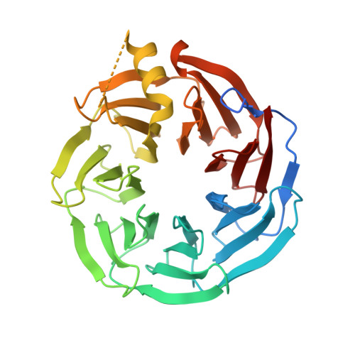
Crystal structure of Human Platelet-activating factor acetylhydrolase IB subunit beta (PAFAH1B1)
Hutchinson, A., Seitova, A., Dong, A., Loppnau, P., Edwards, A.M., Arrowsmith, C.H., Halabelian, L., Structural Genomics Consortium (SGC)To be published.