Structure-Activity Relationship of USP5 Inhibitors.
Mann, M.K., Zepeda-Velazquez, C.A., Gonzalez-Alvarez, H., Dong, A., Kiyota, T., Aman, A.M., Loppnau, P., Li, Y., Wilson, B., Arrowsmith, C.H., Al-Awar, R., Harding, R.J., Schapira, M.(2021) J Med Chem 64: 15017-15036
- PubMed: 34648286 Search on PubMed
- DOI: https://doi.org/10.1021/acs.jmedchem.1c00889
- Primary Citation Related Structures:
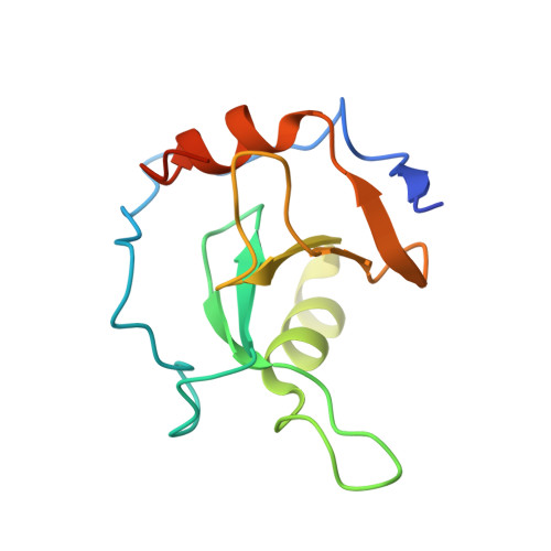
7MS5, 7MS6, 7MS7 - PubMed Abstract:
USP5 is a deubiquitinase that has been implicated in a range of diseases, including cancer, but no USP5-targeting chemical probe has been reported to date. Here, we present the progression of a chemical series that occupies the C-terminal ubiquitin-binding site of a poorly characterized zinc-finger ubiquitin binding domain (ZnF-UBD) of USP5 and competitively inhibits the catalytic activity of the enzyme. Exploration of the structure-activity relationship, complemented with crystallographic characterization of the ZnF-UBD bound to multiple ligands, led to the identification of 64 , which binds to the USP5 ZnF-UBD with a K D of 2.8 μM and is selective over nine proteins containing structurally similar ZnF-UBD domains. 64 inhibits the USP5 catalytic cleavage of a di-ubiquitin substrate in an in vitro assay. This study provides a chemical and structural framework for the discovery of a chemical probe to delineate USP5 function in cells.
- Structural Genomics Consortium, University of Toronto, 101 College Street, MaRS South Tower, Suite 700, Toronto, Ontario M5G 1L7, Canada.
Organizational Affiliation: