7MN4

CmcB E35G mutant from Type II Cut MCP


Experimental Data Snapshot

  • Method: X-RAY DIFFRACTION
  • Resolution: 1.80 Å
  • R-Value Free: 0.240 
  • R-Value Work: 0.200 
  • R-Value Observed: 0.204 

wwPDB Validation   3D Report Full Report


This is version 1.1 of the entry. See complete history


Literature

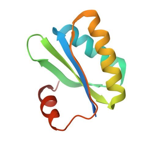
Structural characterization of hexameric shell proteins from two types of choline-utilization bacterial microcompartments

Ochoa, J.M.Mijares, O.Acosta, A.A.Escoto, X.Leon-Rivera, N.Marshall, J.D.Sawaya, M.R.Yeates, T.O.

(2021) Acta Crystallogr F Struct Biol Commun 77: 275-285


Macromolecules
Find similar proteins by:  (by identity cutoff)  |  3D Structure
Entity ID: 1
MoleculeChains Sequence LengthOrganismDetailsImage
BMC domain-containing protein
A, B, C, D, E
A, B, C, D, E, F
101Escherichia coliMutation(s): 2 
Gene Names: pduA_2
UniProt
Find proteins for Q8G9V6 (Escherichia coli)
Explore Q8G9V6 
Go to UniProtKB:  Q8G9V6
Entity Groups  
Sequence Clusters30% Identity50% Identity70% Identity90% Identity95% Identity100% Identity
UniProt GroupQ8G9V6
Sequence Annotations
Expand
  • Reference Sequence
Experimental Data & Validation

Experimental Data

  • Method: X-RAY DIFFRACTION
  • Resolution: 1.80 Å
  • R-Value Free: 0.240 
  • R-Value Work: 0.200 
  • R-Value Observed: 0.204 
  • Space Group: P 1 21 1
Unit Cell:
Length ( Å )Angle ( ˚ )
a = 67.39α = 90
b = 62.72β = 119.95
c = 67.39γ = 90
Software Package:
Software NamePurpose
PHENIXrefinement
XSCALEdata scaling
PHASERphasing
PDB_EXTRACTdata extraction
XDSdata reduction

Structure Validation

View Full Validation Report


Entry History & Funding Information

Deposition Data


Funding OrganizationLocationGrant Number
National Institutes of Health/National Institute Of Allergy and Infectious Diseases (NIH/NIAID)United States--

Revision History  (Full details and data files)

  • Version 1.0: 2021-09-08
    Type: Initial release
  • Version 1.1: 2024-04-03
    Changes: Data collection, Refinement description