7MGY

Sco GlgEI-V279S in complex with 4-alpha-glucoside of valienamine


Experimental Data Snapshot

  • Method: X-RAY DIFFRACTION
  • Resolution: 1.83 Å
  • R-Value Free: 0.217 
  • R-Value Work: 0.185 
  • R-Value Observed: 0.185 

wwPDB Validation   3D Report Full Report


Ligand Structure Quality Assessment 


This is version 1.1 of the entry. See complete history


Literature

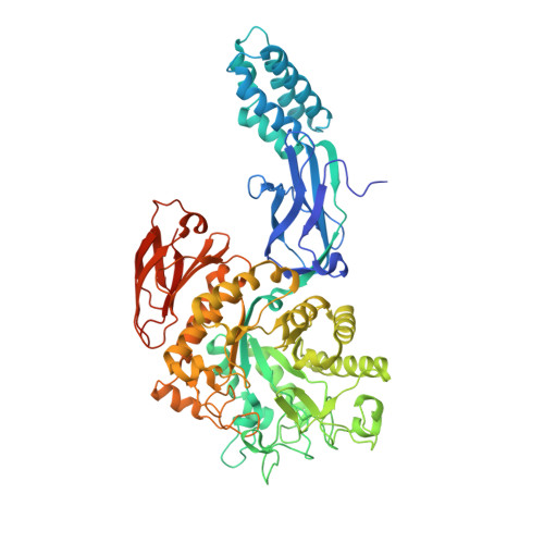
Stereoselective synthesis of a 4-⍺-glucoside of valienamine and its X-ray structure in complex with Streptomyces coelicolor GlgE1-V279S.

Si, A.Jayasinghe, T.D.Thanvi, R.Ronning, D.R.Sucheck, S.J.

(2021) Sci Rep 11: 13413-13413

  • DOI: https://doi.org/10.1038/s41598-021-92554-9
  • Primary Citation of Related Structures:  
    7MEL, 7MGY

  • PubMed Abstract: 

    Glycoside hydrolases (GH) are a large family of hydrolytic enzymes found in all domains of life. As such, they control a plethora of normal and pathogenic biological functions. Thus, understanding selective inhibition of GH enzymes at the atomic level can lead to the identification of new classes of therapeutics. In these studies, we identified a 4-⍺-glucoside of valienamine (8) as an inhibitor of Streptomyces coelicolor (Sco) GlgE1-V279S which belongs to the GH13 Carbohydrate Active EnZyme family. The results obtained from the dose-response experiments show that 8 at a concentration of 1000 µM reduced the enzyme activity of Sco GlgE1-V279S by 65%. The synthetic route to 8 and a closely related 4-⍺-glucoside of validamine (7) was achieved starting from readily available D-maltose. A key step in the synthesis was a chelation-controlled addition of vinylmagnesium bromide to a maltose-derived enone intermediate. X-ray structures of both 7 and 8 in complex with Sco GlgE1-V279S were solved to resolutions of 1.75 and 1.83 Å, respectively. Structural analysis revealed the valienamine derivative 8 binds the enzyme in an E 2 conformation for the cyclohexene fragment. Also, the cyclohexene fragment shows a new hydrogen-bonding contact from the pseudo-diaxial C(3)-OH to the catalytic nucleophile Asp 394 at the enzyme active site. Asp 394, in fact, forms a bidentate interaction with both the C(3)-OH and C(7)-OH of the inhibitor. In contrast, compound 7 disrupts the catalytic sidechain interaction network of Sco GlgE1-V279S via steric interactions resulting in a conformation change in Asp 394. These findings will have implications for the design other aminocarbasugar-based GH13-inhibitors and will be useful for identifying more potent and selective inhibitors.


  • Organizational Affiliation

    Department of Chemistry and Biochemistry, University of Toledo, Toledo, OH, 43606, USA.


Macromolecules
Find similar proteins by:  (by identity cutoff)  |  3D Structure
Entity ID: 1
MoleculeChains Sequence LengthOrganismDetailsImage
Alpha-1,4-glucan:maltose-1-phosphate maltosyltransferase 1
A, B
683Streptomyces coelicolor A3(2)Mutation(s): 1 
Gene Names: glgE1pep1pep1Apep1ISCO5443SC6A11.19c
EC: 2.4.99.16
UniProt
Find proteins for Q9L1K2 (Streptomyces coelicolor (strain ATCC BAA-471 / A3(2) / M145))
Explore Q9L1K2 
Go to UniProtKB:  Q9L1K2
Entity Groups  
Sequence Clusters30% Identity50% Identity70% Identity90% Identity95% Identity100% Identity
UniProt GroupQ9L1K2
Sequence Annotations
Expand
  • Reference Sequence
Small Molecules
Ligands 3 Unique
IDChains Name / Formula / InChI Key2D Diagram3D Interactions
ZD1 (Subject of Investigation/LOI)
Query on ZD1

Download Ideal Coordinates CCD File 
C [auth A],
G [auth B]
(1R,4S,5S,6R)-4-amino-5,6-dihydroxy-2-(hydroxymethyl)cyclohex-2-en-1-yl alpha-D-glucopyranoside
C13 H23 N O9
UINFOZNGFLXUEC-QHWKHWDXSA-N
PGE
Query on PGE

Download Ideal Coordinates CCD File 
D [auth A],
E [auth A],
H [auth B],
I [auth B]
TRIETHYLENE GLYCOL
C6 H14 O4
ZIBGPFATKBEMQZ-UHFFFAOYSA-N
PEG
Query on PEG

Download Ideal Coordinates CCD File 
F [auth A]DI(HYDROXYETHYL)ETHER
C4 H10 O3
MTHSVFCYNBDYFN-UHFFFAOYSA-N
Experimental Data & Validation

Experimental Data

  • Method: X-RAY DIFFRACTION
  • Resolution: 1.83 Å
  • R-Value Free: 0.217 
  • R-Value Work: 0.185 
  • R-Value Observed: 0.185 
  • Space Group: P 41 21 2
Unit Cell:
Length ( Å )Angle ( ˚ )
a = 112.784α = 90
b = 112.784β = 90
c = 310.198γ = 90
Software Package:
Software NamePurpose
PHENIXrefinement
SCALAdata scaling
PDB_EXTRACTdata extraction
MOSFLMdata reduction
PHENIXphasing

Structure Validation

View Full Validation Report


Ligand Structure Quality Assessment 


Entry History & Funding Information

Deposition Data


Funding OrganizationLocationGrant Number
National Institutes of Health/National Institute Of Allergy and Infectious Diseases (NIH/NIAID)United StatesR01AI105084

Revision History  (Full details and data files)

  • Version 1.0: 2021-07-14
    Type: Initial release
  • Version 1.1: 2023-10-18
    Changes: Data collection, Database references, Refinement description