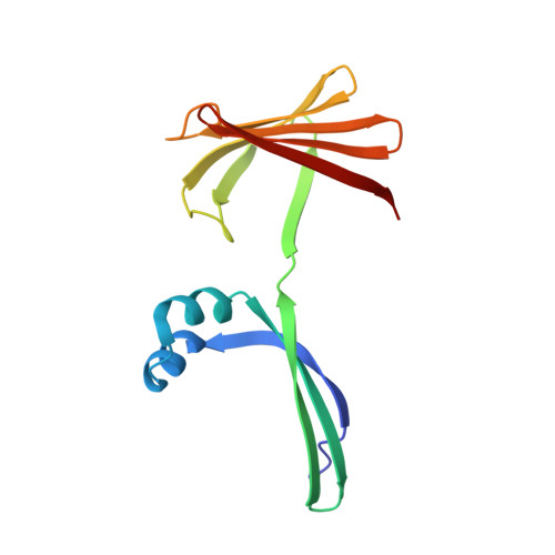
7MFZ

The Crystal Structure of Domain-Swapped Trimer Q108K:K40D:T53A:R58L:Q38F:Q4F Mutant of HCRBPII Bound with LizFluor3 Chromophore Showing Excited State Intermolecular Proton Transfer


Experimental Data Snapshot

  • Method: X-RAY DIFFRACTION
  • Resolution: 2.49 Å
  • R-Value Free: 0.284 
  • R-Value Work: 0.206 
  • R-Value Observed: 0.214 

wwPDB Validation   3D Report Full Report


Ligand Structure Quality Assessment 


This is version 1.1 of the entry. See complete history


Literature

Design of Large Stokes Shift Fluorescent Proteins Based on Excited State Proton Transfer of an Engineered Photobase.

Santos, E.M.Sheng, W.Esmatpour Salmani, R.Tahmasebi Nick, S.Ghanbarpour, A.Gholami, H.Vasileiou, C.Geiger, J.H.Borhan, B.

(2021) J Am Chem Soc 143: 15091-15102

  • DOI: https://doi.org/10.1021/jacs.1c05039
  • Primary Citation of Related Structures:  
    7LSQ, 7MFX, 7MFY, 7MFZ

  • PubMed Abstract: 

    The incredible potential for fluorescent proteins to revolutionize biology has inspired the development of a variety of design strategies to address an equally broad range of photophysical characteristics, depending on potential applications. Of these, fluorescent proteins that simultaneously exhibit high quantum yield, red-shifted emission, and wide separation between excitation and emission wavelengths (Large Stokes Shift, LSS) are rare. The pursuit of LSS systems has led to the formation of a complex, obtained from the marriage of a rationally engineered protein (human cellular retinol binding protein II, hCRBPII) and different fluorogenic molecules, capable of supporting photobase activity. The large increase in basicity upon photoexcitation leads to protonation of the fluorophore in the excited state, dramatically red-shifting its emission, leading to an LSS protein/fluorophore complex. Essential for selective photobase activity is the intimate involvement of the target protein structure and sequence that enables Excited State Proton Transfer (ESPT). The potential power and usefulness of the strategy was demonstrated in live cell imaging of human cell lines.


  • Organizational Affiliation

    Michigan State University, Department of Chemistry, East Lansing, Michigan 48824, United States.


Macromolecules
Find similar proteins by:  (by identity cutoff)  |  3D Structure
Entity ID: 1
MoleculeChains Sequence LengthOrganismDetailsImage
Retinol-binding protein 2
A, B, C
133Homo sapiensMutation(s): 6 
Gene Names: RBP2CRBP2
UniProt & NIH Common Fund Data Resources
Find proteins for P50120 (Homo sapiens)
Explore P50120 
Go to UniProtKB:  P50120
PHAROS:  P50120
GTEx:  ENSG00000114113 
Entity Groups  
Sequence Clusters30% Identity50% Identity70% Identity90% Identity95% Identity100% Identity
UniProt GroupP50120
Sequence Annotations
Expand
  • Reference Sequence
Experimental Data & Validation

Experimental Data

  • Method: X-RAY DIFFRACTION
  • Resolution: 2.49 Å
  • R-Value Free: 0.284 
  • R-Value Work: 0.206 
  • R-Value Observed: 0.214 
  • Space Group: P 2 21 21
Unit Cell:
Length ( Å )Angle ( ˚ )
a = 36.932α = 90
b = 102.41β = 90
c = 108.478γ = 90
Software Package:
Software NamePurpose
PHENIXrefinement
HKL-2000data reduction
HKL-2000data scaling
PHASERphasing

Structure Validation

View Full Validation Report


Ligand Structure Quality Assessment 


Entry History & Funding Information

Deposition Data


Funding OrganizationLocationGrant Number
National Institutes of Health/National Institute of General Medical Sciences (NIH/NIGMS)United StatesGM101353

Revision History  (Full details and data files)

  • Version 1.0: 2022-01-12
    Type: Initial release
  • Version 1.1: 2023-10-18
    Changes: Data collection, Refinement description