7MFO | pdb_00007mfo

X-ray structure of the L136 Aminotransferase from Acanthamoeba polyphaga mimivirus in the presence of TDP and PMP


Experimental Data Snapshot

  • Method: X-RAY DIFFRACTION
  • Resolution: 1.70 Å
  • R-Value Free: 
    0.222 (Depositor), 0.230 (DCC) 
  • R-Value Work: 
    0.182 (Depositor), 0.192 (DCC) 
  • R-Value Observed: 
    0.184 (Depositor) 

wwPDB Validation 3D Report Full Report

Validation slider image for 7MFO

Ligand Structure Quality Assessment 


This is version 1.3 of the entry. See complete history

Literature

Characterization of an aminotransferase from Acanthamoeba polyphaga Mimivirus.

Seltzner, C.A.Ferek, J.D.Thoden, J.B.Holden, H.M.

(2021) Protein Sci 30: 1882-1894

  • DOI: https://doi.org/10.1002/pro.4139
  • Primary Citation Related Structures: 
    7MFO, 7MFP, 7MFQ

  • PubMed Abstract: 

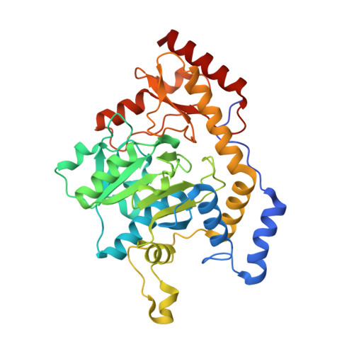
    Acanthamoeba polyphaga Mimivirus, a complex virus that infects amoeba, was first reported in 2003. It is now known that its DNA genome encodes for nearly 1,000 proteins including enzymes that are required for the biosynthesis of the unusual sugar 4-amino-4,6-dideoxy-d-glucose, also known as d-viosamine. As observed in some bacteria, the pathway for the production of this sugar initiates with a nucleotide-linked sugar, which in the Mimivirus is thought to be UDP-d-glucose. The enzyme required for the installment of the amino group at the C-4' position of the pyranosyl moiety is encoded in the Mimivirus by the L136 gene. Here, we describe a structural and functional analysis of this pyridoxal 5'-phosphate-dependent enzyme, referred to as L136. For this analysis, three high-resolution X-ray structures were determined: the wildtype enzyme/pyridoxamine 5'-phosphate/dTDP complex and the site-directed mutant variant K185A in the presence of either UDP-4-amino-4,6-dideoxy-d-glucose or dTDP-4-amino-4,6-dideoxy-d-glucose. Additionally, the kinetic parameters of the enzyme utilizing either UDP-d-glucose or dTDP-d-glucose were measured and demonstrated that L136 is efficient with both substrates. This is in sharp contrast to the structurally related DesI from Streptomyces venezuelae, whose three-dimensional architecture was previously reported by this laboratory. As determined in this investigation, DesI shows a profound preference in its catalytic efficiency for the dTDP-linked sugar substrate. This difference can be explained in part by a hydrophobic patch in DesI that is missing in L136. Notably, the structure of L136 reported here represents the first three-dimensional model for a virally encoded PLP-dependent enzyme and thus provides new information on sugar aminotransferases in general.


  • Organizational Affiliation
    • Department of Biochemistry, University of Wisconsin, Madison, Wisconsin, USA.

Macromolecule Content 

  • Total Structure Weight: 410.02 kDa 
  • Atom Count: 31,417 
  • Modeled Residue Count: 3,508 
  • Deposited Residue Count: 3,560 
  • Unique protein chains: 1

Macromolecules

Find similar proteins by:|  3D Structure
Entity ID: 1
MoleculeChains  Sequence LengthOrganismDetailsImage
L136 aminotransferase
A, B, C, D, E
A, B, C, D, E, F, G, H, I, J
356Acanthamoeba polyphaga mimivirusMutation(s): 0 
Gene Names: MIMI_L136
UniProt
Find proteins for Q5UPL1 (Acanthamoeba polyphaga mimivirus)
Explore Q5UPL1 
Go to UniProtKB:  Q5UPL1
Entity Groups
Sequence Clusters30% Identity50% Identity70% Identity90% Identity95% Identity100% Identity
UniProt GroupQ5UPL1
Sequence Annotations
Expand
Reference Sequence

Small Molecules

Ligands 5 Unique
IDChains Name / Formula / InChI Key2D Diagram3D Interactions
TYD

Query on TYD


Download:Ideal Coordinates CCD File
AA [auth E]
FA [auth G]
GA [auth H]
JA [auth I]
KA [auth J]
AA [auth E],
FA [auth G],
GA [auth H],
JA [auth I],
KA [auth J],
O [auth B],
Q [auth B],
U [auth C],
W [auth D],
Z [auth E]
THYMIDINE-5'-DIPHOSPHATE
C10 H16 N2 O11 P2
UJLXYODCHAELLY-XLPZGREQSA-N
PMP
(Subject of Investigation/LOI)

Query on PMP


Download:Ideal Coordinates CCD File
CA [auth F]
EA [auth G]
HA [auth H]
IA [auth I]
K [auth A]
CA [auth F],
EA [auth G],
HA [auth H],
IA [auth I],
K [auth A],
LA [auth J],
P [auth B],
T [auth C],
X [auth D],
Y [auth E]
4'-DEOXY-4'-AMINOPYRIDOXAL-5'-PHOSPHATE
C8 H13 N2 O5 P
ZMJGSOSNSPKHNH-UHFFFAOYSA-N
PO4

Query on PO4


Download:Ideal Coordinates CCD File
L [auth A]PHOSPHATE ION
O4 P
NBIIXXVUZAFLBC-UHFFFAOYSA-K
EDO

Query on EDO


Download:Ideal Coordinates CCD File
BA [auth E]
DA [auth F]
M [auth A]
MA [auth J]
NA [auth J]
BA [auth E],
DA [auth F],
M [auth A],
MA [auth J],
NA [auth J],
R [auth B],
S [auth B],
V [auth C]
1,2-ETHANEDIOL
C2 H6 O2
LYCAIKOWRPUZTN-UHFFFAOYSA-N
CL

Query on CL


Download:Ideal Coordinates CCD File
N [auth A]CHLORIDE ION
Cl
VEXZGXHMUGYJMC-UHFFFAOYSA-M

Experimental Data & Validation

Experimental Data

  • Method: X-RAY DIFFRACTION
  • Resolution: 1.70 Å
  • R-Value Free:  0.222 (Depositor), 0.230 (DCC) 
  • R-Value Work:  0.182 (Depositor), 0.192 (DCC) 
  • R-Value Observed: 0.184 (Depositor) 
Space Group: P 1 21 1
Unit Cell:
Length ( Å )Angle ( ˚ )
a = 74.885α = 90
b = 126.875β = 90.89
c = 206.365γ = 90
Software Package:
Software NamePurpose
REFMACrefinement
PDB_EXTRACTdata extraction
HKL-3000data reduction
HKL-3000data scaling
PHASERphasing

Structure Validation

View Full Validation Report


Ligand Structure Quality Assessment 


Entry History 

& Funding Information

Deposition Data


Funding OrganizationLocationGrant Number
National Institutes of Health/National Institute of General Medical Sciences (NIH/NIGMS)United StatesR35 GM134643

Revision History  (Full details and data files)

  • Version 1.0: 2021-04-28
    Type: Initial release
  • Version 1.1: 2021-07-14
    Changes: Database references
  • Version 1.2: 2021-09-01
    Changes: Database references
  • Version 1.3: 2024-05-22
    Changes: Data collection