7MF3 | pdb_00007mf3

Structure of the autoinhibited state of smooth muscle myosin-2


Experimental Data Snapshot

  • Method: ELECTRON MICROSCOPY
  • Resolution: 3.40 Å
  • Aggregation State: TISSUE 
  • Reconstruction Method: SINGLE PARTICLE 

Starting Model: experimental
View more details

wwPDB Validation 3D Report Full Report

Validation slider image for 7MF3

This is version 1.1 of the entry. See complete history

Literature

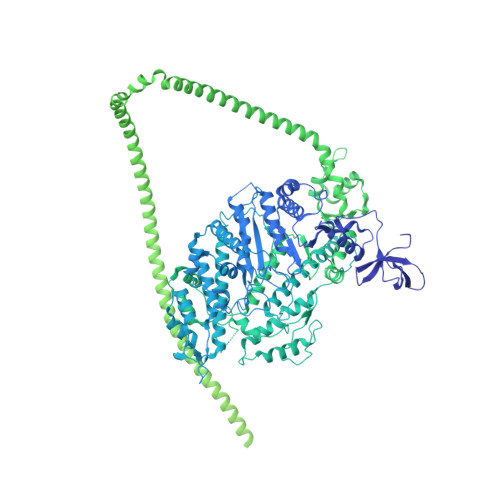
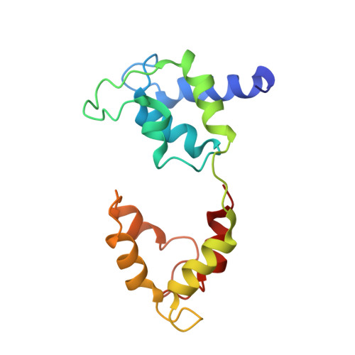
Cryo-EM structure of the autoinhibited state of myosin-2.

Heissler, S.M.Arora, A.S.Billington, N.Sellers, J.R.Chinthalapudi, K.

(2021) Sci Adv 7: eabk3273-eabk3273

  • DOI: https://doi.org/10.1126/sciadv.abk3273
  • Primary Citation Related Structures: 
    7MF3

  • PubMed Abstract: 

    We solved the near-atomic resolution structure of smooth muscle myosin-2 in the autoinhibited state (10 S ) using single-particle cryo–electron microscopy. The 3.4-Å structure reveals the precise molecular architecture of 10 S and the structural basis for myosin-2 regulation. We reveal the position of the phosphorylation sites that control myosin autoinhibition and activation by phosphorylation of the regulatory light chain. Further, we present a previously unidentified conformational state in myosin-2 that traps ADP and P i produced by the hydrolysis of ATP in the active site. This noncanonical state represents a branch of the myosin enzyme cycle and explains the autoinhibition of the enzyme function of 10 S along with its reduced affinity for actin. Together, our structure defines the molecular mechanisms that drive 10 S formation, stabilization, and relief by phosphorylation of the regulatory light chain.


  • Organizational Affiliation
    • Department of Physiology and Cell Biology, Dorothy M. Davis Heart and Lung Research Institute, The Ohio State University College of Medicine, Columbus, OH, USA.

Macromolecule Content 

  • Total Structure Weight: 990.45 kDa 
  • Atom Count: 22,938 
  • Modeled Residue Count: 2,822 
  • Deposited Residue Count: 8,554 
  • Unique protein chains: 3

Macromolecules

Find similar proteins by:|  3D Structure
Entity ID: 1
MoleculeChains  Sequence LengthOrganismDetailsImage
Myosin-11
A, B, G, H
1,978Gallus gallusMutation(s): 0 
UniProt
Find proteins for P10587 (Gallus gallus)
Explore P10587 
Go to UniProtKB:  P10587
Entity Groups
Sequence Clusters30% Identity50% Identity70% Identity90% Identity95% Identity100% Identity
UniProt GroupP10587
Sequence Annotations
Expand
Reference Sequence
Find similar proteins by:|  3D Structure
Entity ID: 2
MoleculeChains  Sequence LengthOrganismDetailsImage
Myosin light polypeptide 6
C, F
150Gallus gallusMutation(s): 0 
UniProt
Find proteins for P02607 (Gallus gallus)
Explore P02607 
Go to UniProtKB:  P02607
Entity Groups
Sequence Clusters30% Identity50% Identity70% Identity90% Identity95% Identity100% Identity
UniProt GroupP02607
Sequence Annotations
Expand
Reference Sequence
Find similar proteins by:|  3D Structure
Entity ID: 3
MoleculeChains  Sequence LengthOrganismDetailsImage
Myosin regulatory light chain 2, smooth muscle major isoform
D, E
171Gallus gallusMutation(s): 0 
UniProt
Find proteins for P02612 (Gallus gallus)
Explore P02612 
Go to UniProtKB:  P02612
Entity Groups
Sequence Clusters30% Identity50% Identity70% Identity90% Identity95% Identity100% Identity
UniProt GroupP02612
Sequence Annotations
Expand
Reference Sequence

Small Molecules

Ligands 3 Unique
IDChains Name / Formula / InChI Key2D Diagram3D Interactions
ADP

Query on ADP


Download:Ideal Coordinates CCD File
I [auth A],
L [auth B]
ADENOSINE-5'-DIPHOSPHATE
C10 H15 N5 O10 P2
XTWYTFMLZFPYCI-KQYNXXCUSA-N
PO4

Query on PO4


Download:Ideal Coordinates CCD File
J [auth A],
M [auth B]
PHOSPHATE ION
O4 P
NBIIXXVUZAFLBC-UHFFFAOYSA-K
MG

Query on MG


Download:Ideal Coordinates CCD File
K [auth A],
N [auth B],
O [auth D],
P [auth E]
MAGNESIUM ION
Mg
JLVVSXFLKOJNIY-UHFFFAOYSA-N

Experimental Data & Validation

Experimental Data

  • Method: ELECTRON MICROSCOPY
  • Resolution: 3.40 Å
  • Aggregation State: TISSUE 
  • Reconstruction Method: SINGLE PARTICLE 
EM Software:
TaskSoftware PackageVersion
RECONSTRUCTIONcryoSPARC
MODEL REFINEMENTPHENIX

Structure Validation

View Full Validation Report


Entry History 

Deposition Data

Revision History  (Full details and data files)

  • Version 1.0: 2022-01-05
    Type: Initial release
  • Version 1.1: 2024-05-29
    Changes: Data collection, Refinement description