7MEZ | pdb_00007mez

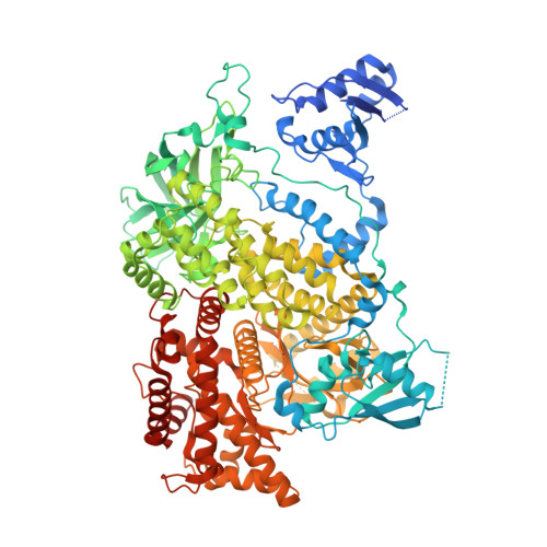
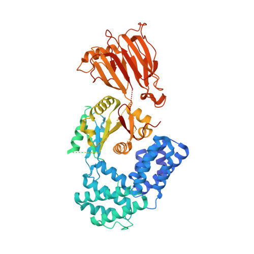
Structure of the phosphoinositide 3-kinase p110 gamma (PIK3CG) p101 (PIK3R5) complex


Experimental Data Snapshot

  • Method: ELECTRON MICROSCOPY
  • Resolution: 2.89 Å
  • Aggregation State: PARTICLE 
  • Reconstruction Method: SINGLE PARTICLE 

wwPDB Validation 3D Report Full Report

Validation slider image for 7MEZ

This is version 1.3 of the entry. See complete history. 

Literature

Structure of the phosphoinositide 3-kinase (PI3K) p110 gamma-p101 complex reveals molecular mechanism of GPCR activation.

Rathinaswamy, M.K., Dalwadi, U., Fleming, K.D., Adams, C., Stariha, J.T.B., Pardon, E., Baek, M., Vadas, O., DiMaio, F., Steyaert, J., Hansen, S.D., Yip, C.K., Burke, J.E.

(2021) Sci Adv 7

  • DOI: https://doi.org/10.1126/sciadv.abj4282
  • Primary Citation Related Structures: 
    7MEZ

  • PubMed Abstract: 

    The class IB phosphoinositide 3-kinase (PI3K), PI3Kγ, is a master regulator of immune cell function and a promising drug target for both cancer and inflammatory diseases. Critical to PI3Kγ function is the association of the p110γ catalytic subunit to either a p101 or p84 regulatory subunit, which mediates activation by G protein-coupled receptors. Here, we report the cryo-electron microscopy structure of a heterodimeric PI3Kγ complex, p110γ-p101. This structure reveals a unique assembly of catalytic and regulatory subunits that is distinct from other class I PI3K complexes. p101 mediates activation through its Gβγ-binding domain, recruiting the heterodimer to the membrane and allowing for engagement of a secondary Gβγ-binding site in p110γ. Mutations at the p110γ-p101 and p110γ-adaptor binding domain interfaces enhanced Gβγ activation. A nanobody that specifically binds to the p101-Gβγ interface blocks activation, providing a novel tool to study and target p110γ-p101-specific signaling events in vivo.


  • Organizational Affiliation: 
    • Department of Biochemistry and Microbiology, University of Victoria, Victoria, British Columbia, Canada.

Macromolecule Content 

  • Total Structure Weight: 224.1 kDa 
  • Atom Count: 12,361 
  • Modeled Residue Count: 1,536 
  • Deposited Residue Count: 1,983 
  • Unique protein chains: 2

Macromolecules

Find similar proteins by:|  3D Structure
Entity ID: 1
MoleculeChains  Sequence LengthOrganismDetailsImage
Phosphatidylinositol 4,5-bisphosphate 3-kinase catalytic subunit gamma isoform1,102Homo sapiensMutation(s): 0 
Gene Names: PIK3CG
EC: 2.7.1.137 (PDB Primary Data), 2.7.1.153 (PDB Primary Data), 2.7.1.154 (PDB Primary Data), 2.7.11.1 (PDB Primary Data)
UniProt & NIH Common Fund Data Resources
Find proteins for P48736 (Homo sapiens)
Explore P48736 
Go to UniProtKB:  P48736
PHAROS:  P48736
GTEx:  ENSG00000105851 
Entity Groups
Sequence Clusters30% Identity50% Identity70% Identity90% Identity95% Identity100% Identity
UniProt GroupP48736
Sequence Annotations
Expand
Reference Sequence
Find similar proteins by:|  3D Structure
Entity ID: 2
MoleculeChains  Sequence LengthOrganismDetailsImage
Phosphoinositide 3-kinase regulatory subunit 5881Sus scrofaMutation(s): 2 
Gene Names: PIK3R5
UniProt
Find proteins for O02696 (Sus scrofa)
Explore O02696 
Go to UniProtKB:  O02696
Entity Groups
Sequence Clusters30% Identity50% Identity70% Identity90% Identity95% Identity100% Identity
UniProt GroupO02696
Sequence Annotations
Expand
Reference Sequence

Experimental Data & Validation

Experimental Data

  • Method: ELECTRON MICROSCOPY
  • Resolution: 2.89 Å
  • Aggregation State: PARTICLE 
  • Reconstruction Method: SINGLE PARTICLE 
EM Software:
TaskSoftware PackageVersion
MODEL REFINEMENTPHENIX
RECONSTRUCTIONcryoSPARC3.0

Structure Validation

View Full Validation Report


Entry History 

& Funding Information

Deposition Data


Funding OrganizationLocationGrant Number
Canadian Institutes of Health Research (CIHR)Canada168998

Revision History  (Full details and data files)

  • Version 1.0: 2021-07-14
    Type: Initial release
  • Version 1.1: 2022-04-06
    Changes: Database references
  • Version 1.2: 2024-05-29
    Changes: Data collection
  • Version 1.3: 2025-05-14
    Changes: Data collection, Structure summary