


Site 2 of the Yersinia pestis substrate-binding protein YfeA is a dynamic surface metal-binding site
Radka, C.D., Aller, S.G.(2021) Acta Crystallogr F Struct Biol Commun 77: 286-293
Experimental Data Snapshot
Starting Model: experimental
View more details
wwPDB Validation 3D Report Full Report

(2021) Acta Crystallogr F Struct Biol Commun 77: 286-293
Entity ID: 1 | |||||
|---|---|---|---|---|---|
| Molecule | Chains | Sequence Length | Organism | Details | Image |
| Periplasmic chelated iron-binding protein YfeA | 323 | Yersinia pestis | Mutation(s): 0 Gene Names: yfeA, YPO2439, y1897, YP_2227 | 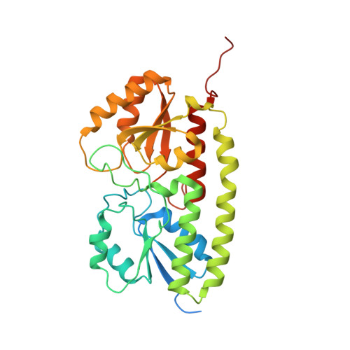 | |
UniProt | |||||
Entity Groups | |||||
| Sequence Clusters | 30% Identity50% Identity70% Identity90% Identity95% Identity100% Identity | ||||
| UniProt Group | Q56952 | ||||
Sequence AnnotationsExpand | |||||
Reference Sequence | |||||
| Ligands 3 Unique | |||||
|---|---|---|---|---|---|
| ID | Chains | Name / Formula / InChI Key | 2D Diagram | 3D Interactions | |
| ZN (Subject of Investigation/LOI) Download:Ideal Coordinates CCD File | D [auth A], H [auth B], L [auth B] | ZINC ION Zn PTFCDOFLOPIGGS-UHFFFAOYSA-N |  | ||
| FE (Subject of Investigation/LOI) Download:Ideal Coordinates CCD File | C [auth A], G [auth B] | FE (III) ION Fe VTLYFUHAOXGGBS-UHFFFAOYSA-N |  | ||
| MN (Subject of Investigation/LOI) Download:Ideal Coordinates CCD File | E [auth A] F [auth A] I [auth B] J [auth B] K [auth B] | MANGANESE (II) ION Mn WAEMQWOKJMHJLA-UHFFFAOYSA-N |  | ||
| Length ( Å ) | Angle ( ˚ ) |
|---|---|
| a = 67.31 | α = 90 |
| b = 76.43 | β = 90 |
| c = 107.36 | γ = 90 |
| Software Name | Purpose |
|---|---|
| PHENIX | refinement |
| PDB_EXTRACT | data extraction |
| HKL-2000 | data reduction |
| SCALEPACK | data scaling |
| PHASER | phasing |


