7M98

ATAD2 bromodomain complexed with histone H4K5ac (res 1-10) ligand


Experimental Data Snapshot

  • Method: X-RAY DIFFRACTION
  • Resolution: 1.60 Å
  • R-Value Free: 0.219 
  • R-Value Work: 0.194 
  • R-Value Observed: 0.195 

wwPDB Validation   3D Report Full Report


This is version 1.2 of the entry. See complete history


Literature

Coordination of Di-Acetylated Histone Ligands by the ATAD2 Bromodomain.

Evans, C.M.Phillips, M.Malone, K.L.Tonelli, M.Cornilescu, G.Cornilescu, C.Holton, S.J.Gorjanacz, M.Wang, L.Carlson, S.Gay, J.C.Nix, J.C.Demeler, B.Markley, J.L.Glass, K.C.

(2021) Int J Mol Sci 22

  • DOI: https://doi.org/10.3390/ijms22179128
  • Primary Citation of Related Structures:  
    7M98

  • PubMed Abstract: 

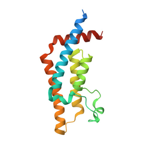
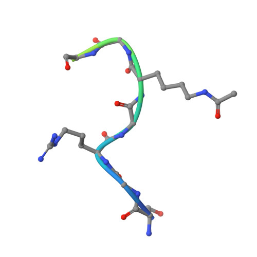
    The ATPase Family, AAA domain-containing protein 2 (ATAD2) bromodomain (BRD) has a canonical bromodomain structure consisting of four α-helices. ATAD2 functions as a co-activator of the androgen and estrogen receptors as well as the MYC and E2F transcription factors. ATAD2 also functions during DNA replication, recognizing newly synthesized histones. In addition, ATAD2 is shown to be up-regulated in multiple forms of cancer including breast, lung, gastric, endometrial, renal, and prostate. Furthermore, up-regulation of ATAD2 is strongly correlated with poor prognosis in many types of cancer, making the ATAD2 bromodomain an innovative target for cancer therapeutics. In this study, we describe the recognition of histone acetyllysine modifications by the ATAD2 bromodomain. Residue-specific information on the complex formed between the histone tail and the ATAD2 bromodomain, obtained through nuclear magnetic resonance spectroscopy (NMR) and X-ray crystallography, illustrates key residues lining the binding pocket, which are involved in coordination of di-acetylated histone tails. Analytical ultracentrifugation, NMR relaxation data, and isothermal titration calorimetry further confirm the monomeric state of the functionally active ATAD2 bromodomain in complex with di-acetylated histone ligands. Overall, we describe histone tail recognition by ATAD2 BRD and illustrate that one acetyllysine group is primarily engaged by the conserved asparagine (N1064), the "RVF" shelf residues, and the flexible ZA loop. Coordination of a second acetyllysine group also occurs within the same binding pocket but is essentially governed by unique hydrophobic and electrostatic interactions making the di-acetyllysine histone coordination more specific than previously presumed.


  • Organizational Affiliation

    Department of Pharmaceutical Sciences, Albany College of Pharmacy and Health Sciences, Colchester, VT 05446, USA.


Macromolecules
Find similar proteins by:  (by identity cutoff)  |  3D Structure
Entity ID: 1
MoleculeChains Sequence LengthOrganismDetailsImage
ATPase family AAA domain-containing protein 2152Homo sapiensMutation(s): 1 
Gene Names: ATAD2L16PRO2000
EC: 3.6.1
UniProt & NIH Common Fund Data Resources
Find proteins for Q6PL18 (Homo sapiens)
Explore Q6PL18 
Go to UniProtKB:  Q6PL18
PHAROS:  Q6PL18
GTEx:  ENSG00000156802 
Entity Groups  
Sequence Clusters30% Identity50% Identity70% Identity90% Identity95% Identity100% Identity
UniProt GroupQ6PL18
Sequence Annotations
Expand
  • Reference Sequence

Find similar proteins by:  Sequence   |   3D Structure  

Entity ID: 2
MoleculeChains Sequence LengthOrganismDetailsImage
Histone H415Homo sapiensMutation(s): 0 
Gene Names: 
UniProt & NIH Common Fund Data Resources
Find proteins for P62805 (Homo sapiens)
Explore P62805 
Go to UniProtKB:  P62805
PHAROS:  P62805
Entity Groups  
Sequence Clusters30% Identity50% Identity70% Identity90% Identity95% Identity100% Identity
UniProt GroupP62805
Sequence Annotations
Expand
  • Reference Sequence
Small Molecules
Modified Residues  1 Unique
IDChains TypeFormula2D DiagramParent
ALY
Query on ALY
B
L-PEPTIDE LINKINGC8 H16 N2 O3LYS
Experimental Data & Validation

Experimental Data

  • Method: X-RAY DIFFRACTION
  • Resolution: 1.60 Å
  • R-Value Free: 0.219 
  • R-Value Work: 0.194 
  • R-Value Observed: 0.195 
  • Space Group: P 43 2 2
Unit Cell:
Length ( Å )Angle ( ˚ )
a = 53.56α = 90
b = 53.56β = 90
c = 160.35γ = 90
Software Package:
Software NamePurpose
PHENIXrefinement
PHENIXrefinement
Cootmodel building
XDSdata reduction
XDSdata scaling
PHASERphasing

Structure Validation

View Full Validation Report


Entry History & Funding Information

Deposition Data


Funding OrganizationLocationGrant Number
National Institutes of Health/National Institute of General Medical Sciences (NIH/NIGMS)United StatesR01GM129338
National Institutes of Health/National Institute of General Medical Sciences (NIH/NIGMS)United StatesR15GM104865

Revision History  (Full details and data files)

  • Version 1.0: 2021-09-22
    Type: Initial release
  • Version 1.1: 2023-10-18
    Changes: Data collection, Refinement description
  • Version 1.2: 2023-11-15
    Changes: Data collection