7M96

Bovine sigma-2 receptor bound to Z4857158944


Experimental Data Snapshot

  • Method: X-RAY DIFFRACTION
  • Resolution: 2.41 Å
  • R-Value Free: 0.288 
  • R-Value Work: 0.250 
  • R-Value Observed: 0.252 

wwPDB Validation   3D Report Full Report


Ligand Structure Quality Assessment 


This is version 1.3 of the entry. See complete history


Literature

Structures of the sigma 2 receptor enable docking for bioactive ligand discovery.

Alon, A.Lyu, J.Braz, J.M.Tummino, T.A.Craik, V.O'Meara, M.J.Webb, C.M.Radchenko, D.S.Moroz, Y.S.Huang, X.P.Liu, Y.Roth, B.L.Irwin, J.J.Basbaum, A.I.Shoichet, B.K.Kruse, A.C.

(2021) Nature 600: 759-764

  • DOI: https://doi.org/10.1038/s41586-021-04175-x
  • Primary Citation of Related Structures:  
    7M93, 7M94, 7M95, 7M96, 7MFI

  • PubMed Abstract: 

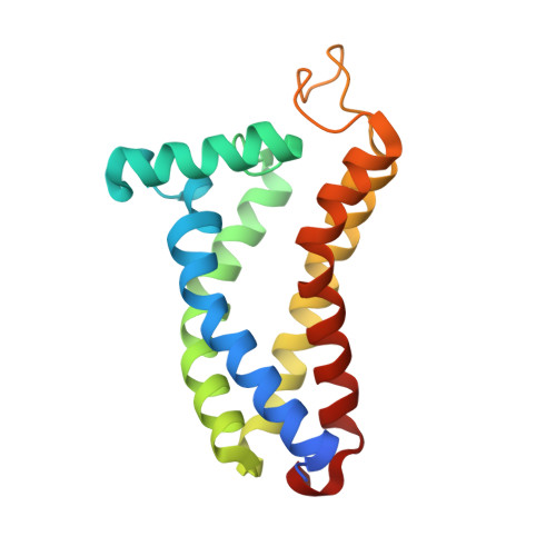
    The σ 2 receptor has attracted intense interest in cancer imaging 1 , psychiatric disease 2 , neuropathic pain 3-5 and other areas of biology 6,7 . Here we determined the crystal structure of this receptor in complex with the clinical candidate roluperidone 2 and the tool compound PB28 8 . These structures templated a large-scale docking screen of 490 million virtual molecules, of which 484 compounds were synthesized and tested. We identified 127 new chemotypes with affinities superior to 1 μM, 31 of which had affinities superior to 50 nM. The hit rate fell smoothly and monotonically with docking score. We optimized three hits for potency and selectivity, and achieved affinities that ranged from 3 to 48 nM, with up to 250-fold selectivity versus the σ 1 receptor. Crystal structures of two ligands bound to the σ 2 receptor confirmed the docked poses. To investigate the contribution of the σ 2 receptor in pain, two potent σ 2 -selective ligands and one potent σ 1 2 non-selective ligand were tested for efficacy in a mouse model of neuropathic pain. All three ligands showed time-dependent decreases in mechanical hypersensitivity in the spared nerve injury model 9 , suggesting that the σ 2 receptor has a role in nociception. This study illustrates the opportunities for rapid discovery of in vivo probes through structure-based screens of ultra large libraries, enabling study of underexplored areas of biology.


  • Organizational Affiliation

    Department of Biological Chemistry and Molecular Pharmacology, Blavatnik Institute, Harvard Medical School, Boston, MA, USA.


Macromolecules
Find similar proteins by:  (by identity cutoff)  |  3D Structure
Entity ID: 1
MoleculeChains Sequence LengthOrganismDetailsImage
Sigma intracellular receptor 2
A, B, C, D
174Bos taurusMutation(s): 0 
Gene Names: TMEM97S2R
Membrane Entity: Yes 
UniProt
Find proteins for Q3MHW7 (Bos taurus)
Explore Q3MHW7 
Go to UniProtKB:  Q3MHW7
Entity Groups  
Sequence Clusters30% Identity50% Identity70% Identity90% Identity95% Identity100% Identity
UniProt GroupQ3MHW7
Sequence Annotations
Expand
  • Reference Sequence
Small Molecules
Ligands 3 Unique
IDChains Name / Formula / InChI Key2D Diagram3D Interactions
OLC
Query on OLC

Download Ideal Coordinates CCD File 
AA [auth D]
BA [auth D]
CA [auth D]
F [auth A]
G [auth A]
AA [auth D],
BA [auth D],
CA [auth D],
F [auth A],
G [auth A],
H [auth A],
I [auth A],
J [auth A],
K [auth A],
M [auth A],
O [auth B],
P [auth B],
Q [auth B],
R [auth B],
S [auth B],
U [auth C],
V [auth C],
W [auth C],
Y [auth D],
Z [auth D]
(2R)-2,3-dihydroxypropyl (9Z)-octadec-9-enoate
C21 H40 O4
RZRNAYUHWVFMIP-GDCKJWNLSA-N
YTD (Subject of Investigation/LOI)
Query on YTD

Download Ideal Coordinates CCD File 
E [auth A],
N [auth B],
T [auth C],
X [auth D]
6-({[(1S)-1-hydroxy-2-methyl-1-phenylpropan-2-yl]amino}methyl)-1-methyl-3,4-dihydroquinolin-2(1H)-one
C21 H26 N2 O2
QITHCLWWRWXBDJ-FQEVSTJZSA-N
MES
Query on MES

Download Ideal Coordinates CCD File 
DA [auth D],
L [auth A]
2-(N-MORPHOLINO)-ETHANESULFONIC ACID
C6 H13 N O4 S
SXGZJKUKBWWHRA-UHFFFAOYSA-N
Experimental Data & Validation

Experimental Data

  • Method: X-RAY DIFFRACTION
  • Resolution: 2.41 Å
  • R-Value Free: 0.288 
  • R-Value Work: 0.250 
  • R-Value Observed: 0.252 
  • Space Group: P 1 21 1
Unit Cell:
Length ( Å )Angle ( ˚ )
a = 70.7α = 90
b = 55.35β = 94.5
c = 92.95γ = 90
Software Package:
Software NamePurpose
Blu-Icedata collection
XDSdata reduction
XDSdata scaling
Cootmodel building
PHENIXrefinement
PHASERphasing

Structure Validation

View Full Validation Report


Ligand Structure Quality Assessment 


Entry History & Funding Information

Deposition Data


Funding OrganizationLocationGrant Number
National Institutes of Health/National Institute of General Medical Sciences (NIH/NIGMS)United StatesR01GM119185

Revision History  (Full details and data files)

  • Version 1.0: 2021-12-15
    Type: Initial release
  • Version 1.1: 2021-12-22
    Changes: Database references
  • Version 1.2: 2022-01-05
    Changes: Database references
  • Version 1.3: 2023-10-18
    Changes: Data collection, Refinement description