Discovery of IACS-9779 and IACS-70465 as Potent Inhibitors Targeting Indoleamine 2,3-Dioxygenase 1 (IDO1) Apoenzyme.
Hamilton, M.M., Mseeh, F., McAfoos, T.J., Leonard, P.G., Reyna, N.J., Harris, A.L., Xu, A., Han, M., Soth, M.J., Czako, B., Theroff, J.P., Mandal, P.K., Burke, J.P., Virgin-Downey, B., Petrocchi, A., Pfaffinger, D., Rogers, N.E., Parker, C.A., Yu, S.S., Jiang, Y., Krapp, S., Lammens, A., Trevitt, G., Tremblay, M.R., Mikule, K., Wilcoxen, K., Cross, J.B., Jones, P., Marszalek, J.R., Lewis, R.T.(2021) J Med Chem 64: 11302-11329
- PubMed: 34292726 Search on PubMed
- DOI: https://doi.org/10.1021/acs.jmedchem.1c00679
- Primary Citation Related Structures:
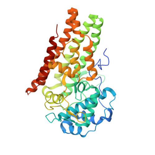
7B1O, 7M63, 7M7D - PubMed Abstract:
Indoleamine 2,3-dioxygenase 1 (IDO1), a heme-containing enzyme that mediates the rate-limiting step in the metabolism of l-tryptophan to kynurenine, has been widely explored as a potential immunotherapeutic target in oncology. We developed a class of inhibitors with a conformationally constrained bicyclo[3.1.0]hexane core. These potently inhibited IDO1 in a cellular context by binding to the apoenzyme, as elucidated by biochemical characterization and X-ray crystallography. A SKOV3 tumor model was instrumental in differentiating compounds, leading to the identification of IACS-9779 ( 62 ) and IACS-70465 ( 71 ). IACS-70465 has excellent cellular potency, a robust pharmacodynamic response, and in a human whole blood assay was more potent than linrodostat (BMS-986205). IACS-9779 with a predicted human efficacious once daily dose below 1 mg/kg to sustain >90% inhibition of IDO1 displayed an acceptable safety margin in rodent toxicology and dog cardiovascular studies to support advancement into preclinical safety evaluation for human development.
- IACS (Institute for Applied Cancer Science), University of Texas, MD Anderson Cancer Center, 1881 East Road, Houston, Texas 77054, United States.
Organizational Affiliation: