7M5W | pdb_00007m5w

Crystal structure of the HMG-C1 domain of human capicua bound to DNA


Experimental Data Snapshot

  • Method: X-RAY DIFFRACTION
  • Resolution: 2.95 Å
  • R-Value Free: 
    0.243 (Depositor), 0.240 (DCC) 
  • R-Value Work: 
    0.209 (Depositor), 0.210 (DCC) 
  • R-Value Observed: 
    0.211 (Depositor) 

Starting Model: experimental
View more details

wwPDB Validation   3D Report Full Report


This is version 1.2 of the entry. See complete history


Literature

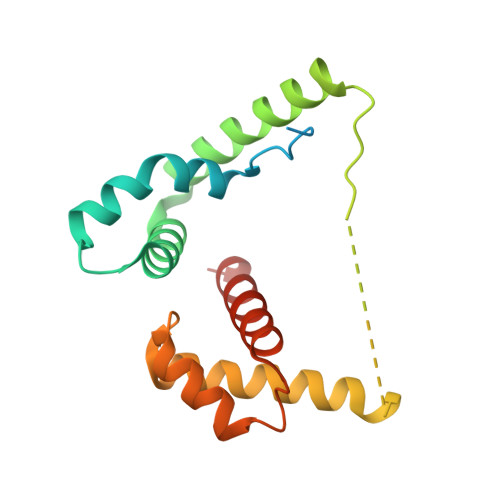
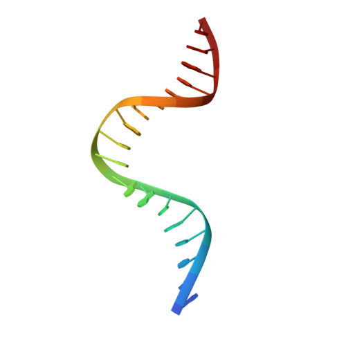
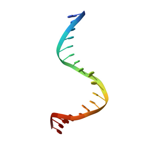
Molecular basis of DNA recognition by the HMG-box-C1 module of capicua.

Webb, J.Liew, J.J.M.Gnann, A.D.Ilkhani, K.Patterson, M.Paul, S.Fores, M.Jimenez, G.Veraksa, A.Dowling, D.P.

(2025) Structure 

  • DOI: https://doi.org/10.1016/j.str.2025.08.018
  • Primary Citation of Related Structures:  
    7M5W

  • PubMed Abstract: 

    The HMG-box protein capicua (CIC) is a conserved transcriptional repressor with key functions in development and disease. CIC binding of DNA requires both its HMG-box and a separate domain called C1. How these domains cooperate to recognize specific DNA sequences is not known. Here, we report the crystal structure of the human CIC HMG-box and C1 domains complexed with a DNA oligomer containing a consensus octameric binding site. We find that both domains adopt tri-helical structures that pack against opposite sides of the DNA helix. The C1 domain folds into a helix-turn-helix (HTH) structure, inserting into the DNA major groove to enhance affinity. We investigate the system using molecular dynamics simulations and binding assays that interrogate the observed HMG-box and C1 domain interface and prominent cancer variants. Our results reveal a unique bipartite DNA-binding module and provide insights into the effects of cancer and domain interface mutations.


  • Organizational Affiliation
    • Chemistry Department, University of Massachusetts Boston, Boston, MA 02125, USA.

Macromolecules

Find similar proteins by:  (by identity cutoff)  |  3D Structure
Entity ID: 1
MoleculeChains Sequence LengthOrganismDetailsImage
Protein capicua homolog186Homo sapiensMutation(s): 0 
Gene Names: CICKIAA0306
UniProt & NIH Common Fund Data Resources
Find proteins for Q96RK0 (Homo sapiens)
Explore Q96RK0 
Go to UniProtKB:  Q96RK0
PHAROS:  Q96RK0
GTEx:  ENSG00000079432 
Entity Groups  
Sequence Clusters30% Identity50% Identity70% Identity90% Identity95% Identity100% Identity
UniProt GroupQ96RK0
Sequence Annotations
Expand
  • Reference Sequence

Find similar nucleic acids by:  Sequence   |   3D Structure  

Entity ID: 2
MoleculeChains LengthOrganismImage
DNA (5'-D(*GP*GP*TP*TP*AP*TP*GP*AP*AP*TP*GP*AP*AP*AP*AP*AP*GP*C)-3')18synthetic construct
Sequence Annotations
Expand
  • Reference Sequence

Find similar nucleic acids by:  Sequence   |   3D Structure  

Entity ID: 3
MoleculeChains LengthOrganismImage
DNA (5'-D(*GP*CP*TP*TP*TP*TP*TP*CP*AP*TP*TP*CP*AP*TP*AP*AP*CP*C)-3')18synthetic construct
Sequence Annotations
Expand
  • Reference Sequence
Small Molecules
Ligands 1 Unique
IDChains Name / Formula / InChI Key2D Diagram3D Interactions
CA
Query on CA

Download Ideal Coordinates CCD File 
D [auth B]CALCIUM ION
Ca
BHPQYMZQTOCNFJ-UHFFFAOYSA-N
Experimental Data & Validation

Experimental Data

  • Method: X-RAY DIFFRACTION
  • Resolution: 2.95 Å
  • R-Value Free:  0.243 (Depositor), 0.240 (DCC) 
  • R-Value Work:  0.209 (Depositor), 0.210 (DCC) 
  • R-Value Observed: 0.211 (Depositor) 
Space Group: P 21 21 21
Unit Cell:
Length ( Å )Angle ( ˚ )
a = 46.89α = 90
b = 79.92β = 90
c = 106.96γ = 90
Software Package:
Software NamePurpose
PHENIXrefinement
XDSdata reduction
XSCALEdata scaling
PHASERphasing
PDB_EXTRACTdata extraction

Structure Validation

View Full Validation Report


Entry History 

Deposition Data

Revision History  (Full details and data files)

  • Version 1.0: 2022-04-06
    Type: Initial release
  • Version 1.1: 2023-10-18
    Changes: Data collection, Database references, Refinement description
  • Version 1.2: 2025-11-05
    Changes: Database references, Structure summary