7M5C | pdb_00007m5c

Crystal Structure of human BAK in complex with WT BAK BH3 peptide


Experimental Data Snapshot

  • Method: X-RAY DIFFRACTION
  • Resolution: 3.06 Å
  • R-Value Free: 
    0.249 (Depositor), 0.250 (DCC) 
  • R-Value Work: 
    0.213 (Depositor), 0.214 (DCC) 
  • R-Value Observed: 
    0.216 (Depositor) 

Starting Model: experimental
View more details

wwPDB Validation 3D Report Full Report

Validation slider image for 7M5C

This is version 1.3 of the entry. See complete history

Literature

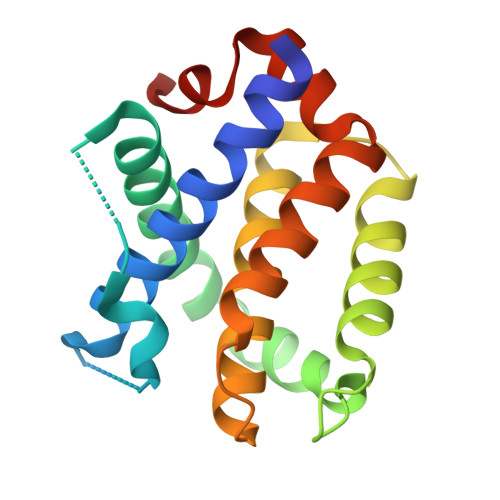
Structural basis of BAK activation in mitochondrial apoptosis initiation.

Singh, G.Guibao, C.D.Seetharaman, J.Aggarwal, A.Grace, C.R.McNamara, D.E.Vaithiyalingam, S.Waddell, M.B.Moldoveanu, T.

(2022) Nat Commun 13: 250-250

  • DOI: https://doi.org/10.1038/s41467-021-27851-y
  • Primary Citation Related Structures: 
    7M5A, 7M5B, 7M5C

  • PubMed Abstract: 

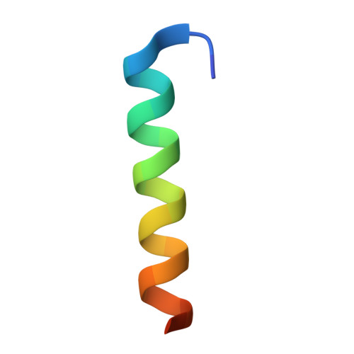
    BCL-2 proteins regulate mitochondrial poration in apoptosis initiation. How the pore-forming BCL-2 Effector BAK is activated remains incompletely understood mechanistically. Here we investigate autoactivation and direct activation by BH3-only proteins, which cooperate to lower BAK threshold in membrane poration and apoptosis initiation. We define in trans BAK autoactivation as the asymmetric "BH3-in-groove" triggering of dormant BAK by active BAK. BAK autoactivation is mechanistically similar to direct activation. The structure of autoactivated BAK BH3-BAK complex reveals the conformational changes leading to helix α1 destabilization, which is a hallmark of BAK activation. Helix α1 is destabilized and restabilized in structures of BAK engaged by rationally designed, high-affinity activating and inactivating BID-like BH3 ligands, respectively. Altogether our data support the long-standing hit-and-run mechanism of BAK activation by transient binding of BH3-only proteins, demonstrating that BH3-induced structural changes are more important in BAK activation than BH3 ligand affinity.


  • Organizational Affiliation
    • Department of Structural Biology, St. Jude Children's Research Hospital, Memphis, TN, USA.

Macromolecule Content 

  • Total Structure Weight: 215.04 kDa 
  • Atom Count: 13,528 
  • Modeled Residue Count: 1,688 
  • Deposited Residue Count: 1,910 
  • Unique protein chains: 2

Macromolecules

Find similar proteins by:|  3D Structure
Entity ID: 1
MoleculeChains  Sequence LengthOrganismDetailsImage
Bcl-2 homologous antagonist/killer
A, C, E, G, I
A, C, E, G, I, K, M, O, Q, S
166Homo sapiensMutation(s): 0 
Gene Names: BAK1BAKBCL2L7CDN1
UniProt & NIH Common Fund Data Resources
Find proteins for Q16611 (Homo sapiens)
Explore Q16611 
Go to UniProtKB:  Q16611
PHAROS:  Q16611
GTEx:  ENSG00000030110 
Entity Groups
Sequence Clusters30% Identity50% Identity70% Identity90% Identity95% Identity100% Identity
UniProt GroupQ16611
Sequence Annotations
Expand
Reference Sequence
Find similar proteins by:  Sequence   |   3D Structure  
Entity ID: 2
MoleculeChains  Sequence LengthOrganismDetailsImage
Bcl-2 homologous antagonist/killer
B, D, F, H, J
B, D, F, H, J, L, N, P, R, T
25Homo sapiensMutation(s): 0 
UniProt & NIH Common Fund Data Resources
Find proteins for Q16611 (Homo sapiens)
Explore Q16611 
Go to UniProtKB:  Q16611
PHAROS:  Q16611
GTEx:  ENSG00000030110 
Entity Groups
Sequence Clusters30% Identity50% Identity70% Identity90% Identity95% Identity100% Identity
UniProt GroupQ16611
Sequence Annotations
Expand
Reference Sequence

Small Molecules

Ligands 2 Unique
IDChains Name / Formula / InChI Key2D Diagram3D Interactions
SO4

Query on SO4


Download:Ideal Coordinates CCD File
AA [auth H]
BA [auth H]
EA [auth M]
FA [auth M]
HA [auth P]
AA [auth H],
BA [auth H],
EA [auth M],
FA [auth M],
HA [auth P],
KA [auth T],
V [auth A],
Z [auth G]
SULFATE ION
O4 S
QAOWNCQODCNURD-UHFFFAOYSA-L
CU

Query on CU


Download:Ideal Coordinates CCD File
CA [auth I]
DA [auth M]
GA [auth O]
IA [auth Q]
JA [auth S]
CA [auth I],
DA [auth M],
GA [auth O],
IA [auth Q],
JA [auth S],
U [auth A],
W [auth C],
X [auth E],
Y [auth G]
COPPER (II) ION
Cu
JPVYNHNXODAKFH-UHFFFAOYSA-N

Experimental Data & Validation

Experimental Data

  • Method: X-RAY DIFFRACTION
  • Resolution: 3.06 Å
  • R-Value Free:  0.249 (Depositor), 0.250 (DCC) 
  • R-Value Work:  0.213 (Depositor), 0.214 (DCC) 
  • R-Value Observed: 0.216 (Depositor) 
Space Group: P 1 21 1
Unit Cell:
Length ( Å )Angle ( ˚ )
a = 89.385α = 90
b = 132.215β = 119.2
c = 89.379γ = 90
Software Package:
Software NamePurpose
PHENIXrefinement
Aimlessdata scaling
PDB_EXTRACTdata extraction
PHASERphasing
XDSdata reduction

Structure Validation

View Full Validation Report


Entry History 

Deposition Data

Revision History  (Full details and data files)

  • Version 1.0: 2022-01-12
    Type: Initial release
  • Version 1.1: 2022-08-03
    Changes: Database references
  • Version 1.2: 2023-10-18
    Changes: Data collection, Refinement description
  • Version 1.3: 2024-10-09
    Changes: Structure summary