7M4E

DNA Polymerase Lambda, dCTP:At Mg2+ Reaction State Ternary Complex, 120 min


Experimental Data Snapshot

  • Method: X-RAY DIFFRACTION
  • Resolution: 1.90 Å
  • R-Value Free: 0.233 
  • R-Value Work: 0.209 
  • R-Value Observed: 0.210 

wwPDB Validation   3D Report Full Report


Ligand Structure Quality Assessment 


This is version 1.1 of the entry. See complete history


Literature

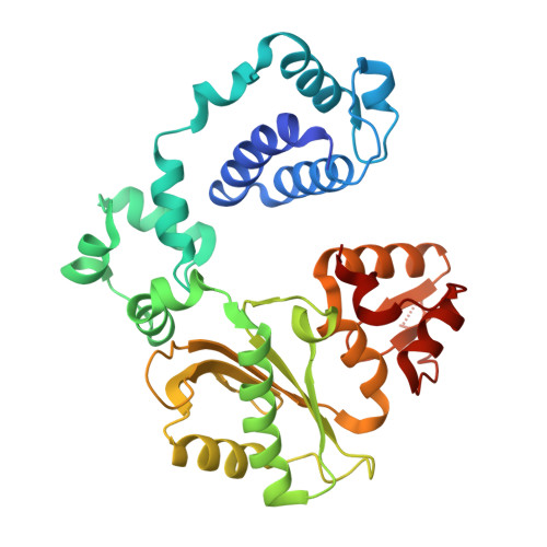
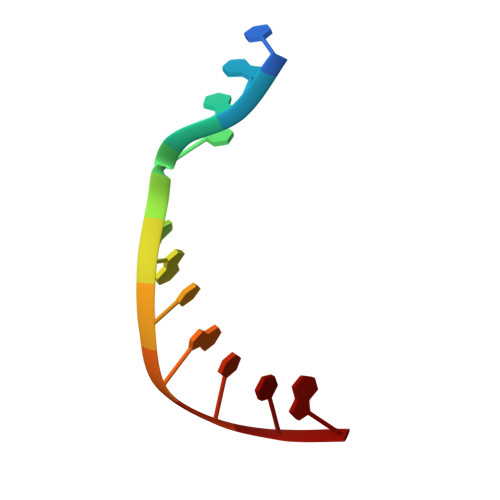
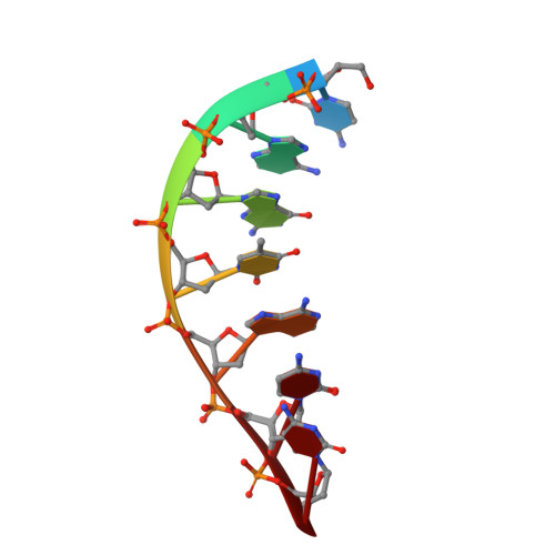
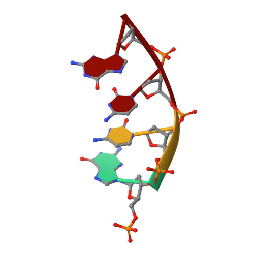
Watching right and wrong nucleotide insertion captures hidden polymerase fidelity checkpoints.

Jamsen, J.A.Shock, D.D.Wilson, S.H.

(2022) Nat Commun 13: 3193-3193

  • DOI: https://doi.org/10.1038/s41467-022-30141-w
  • Primary Citation of Related Structures:  
    7M43, 7M44, 7M45, 7M46, 7M47, 7M48, 7M49, 7M4A, 7M4B, 7M4C, 7M4D, 7M4E, 7M4F, 7M4G, 7M4H, 7M4I, 7M4J, 7M4K, 7M4L

  • PubMed Abstract: 

    Efficient and accurate DNA synthesis is enabled by DNA polymerase fidelity checkpoints that promote insertion of the right instead of wrong nucleotide. Erroneous X-family polymerase (pol) λ nucleotide insertion leads to genomic instability in double strand break and base-excision repair. Here, time-lapse crystallography captures intermediate catalytic states of pol λ undergoing right and wrong natural nucleotide insertion. The revealed nucleotide sensing mechanism responds to base pair geometry through active site deformation to regulate global polymerase-substrate complex alignment in support of distinct optimal (right) or suboptimal (wrong) reaction pathways. An induced fit during wrong but not right insertion, and associated metal, substrate, side chain and pyrophosphate reaction dynamics modulated nucleotide insertion. A third active site metal hastened right but not wrong insertion and was not essential for DNA synthesis. The previously hidden fidelity checkpoints uncovered reveal fundamental strategies of polymerase DNA repair synthesis in genomic instability.


  • Organizational Affiliation

    Genome Integrity and Structural Biology Laboratory, National Institute of Environmental Health Sciences, National Institutes of Health, Research Triangle Park, NC, 27709, USA. joonas.jamsen@nih.gov.


Macromolecules

Find similar proteins by:  (by identity cutoff)  |  3D Structure
Entity ID: 1
MoleculeChains Sequence LengthOrganismDetailsImage
DNA polymerase lambda329Homo sapiensMutation(s): 0 
Gene Names: POLL
EC: 2.7.7.7 (PDB Primary Data), 4.2.99 (PDB Primary Data)
UniProt & NIH Common Fund Data Resources
Find proteins for Q9UGP5 (Homo sapiens)
Explore Q9UGP5 
Go to UniProtKB:  Q9UGP5
PHAROS:  Q9UGP5
GTEx:  ENSG00000166169 
Entity Groups  
Sequence Clusters30% Identity50% Identity70% Identity90% Identity95% Identity100% Identity
UniProt GroupQ9UGP5
Sequence Annotations
Expand
  • Reference Sequence

Find similar nucleic acids by:  Sequence   |   3D Structure  

Entity ID: 2
MoleculeChains LengthOrganismImage
DNA (5'-D(*CP*GP*GP*CP*AP*GP*TP*AP*CP*TP*G)-3')B [auth T]11Homo sapiens
Sequence Annotations
Expand
  • Reference Sequence

Find similar nucleic acids by:  Sequence   |   3D Structure  

Entity ID: 3
MoleculeChains LengthOrganismImage
DNA (5'-D(*CP*AP*GP*TP*AP*CP*C)-3')C [auth P]7Homo sapiens
Sequence Annotations
Expand
  • Reference Sequence

Find similar nucleic acids by:  Sequence   |   3D Structure  

Entity ID: 4
MoleculeChains LengthOrganismImage
DNA (5'-D(P*GP*CP*CP*G)-3')4Homo sapiens
Sequence Annotations
Expand
  • Reference Sequence
Small Molecules
Ligands 5 Unique
IDChains Name / Formula / InChI Key2D Diagram3D Interactions
DCP (Subject of Investigation/LOI)
Query on DCP

Download Ideal Coordinates CCD File 
J [auth A]2'-DEOXYCYTIDINE-5'-TRIPHOSPHATE
C9 H16 N3 O13 P3
RGWHQCVHVJXOKC-SHYZEUOFSA-N
PPV (Subject of Investigation/LOI)
Query on PPV

Download Ideal Coordinates CCD File 
E [auth A]PYROPHOSPHATE
H4 O7 P2
XPPKVPWEQAFLFU-UHFFFAOYSA-N
EDO
Query on EDO

Download Ideal Coordinates CCD File 
K [auth A]1,2-ETHANEDIOL
C2 H6 O2
LYCAIKOWRPUZTN-UHFFFAOYSA-N
MG (Subject of Investigation/LOI)
Query on MG

Download Ideal Coordinates CCD File 
F [auth A],
G [auth A]
MAGNESIUM ION
Mg
JLVVSXFLKOJNIY-UHFFFAOYSA-N
NA
Query on NA

Download Ideal Coordinates CCD File 
H [auth A],
I [auth A]
SODIUM ION
Na
FKNQFGJONOIPTF-UHFFFAOYSA-N
Experimental Data & Validation

Experimental Data

  • Method: X-RAY DIFFRACTION
  • Resolution: 1.90 Å
  • R-Value Free: 0.233 
  • R-Value Work: 0.209 
  • R-Value Observed: 0.210 
  • Space Group: P 21 21 21
Unit Cell:
Length ( Å )Angle ( ˚ )
a = 55.825α = 90
b = 62.088β = 90
c = 142.096γ = 90
Software Package:
Software NamePurpose
PHENIXrefinement
HKL-2000data reduction
HKL-2000data scaling
PDB_EXTRACTdata extraction
HKL-3000phasing

Structure Validation

View Full Validation Report


Ligand Structure Quality Assessment 


Entry History & Funding Information

Deposition Data


Funding OrganizationLocationGrant Number
National Institutes of Health/National Institute of Environmental Health Sciences (NIH/NIEHS)United StatesZ01-ES050158
National Institutes of Health/National Institute of Environmental Health Sciences (NIH/NIEHS)United StatesZ01-ES050161
National Institutes of Health/National Institute of Environmental Health Sciences (NIH/NIEHS)United States1K99ES029572-01

Revision History  (Full details and data files)

  • Version 1.0: 2022-07-06
    Type: Initial release
  • Version 1.1: 2023-10-18
    Changes: Data collection, Refinement description