


B-FACTOR ANALYSIS SUGGEST THAT L-LYSINE AND R, R-BISLYSINE ALLOSTERICALLY INHIBIT Cj.DHDPS ENZYME BY DECREASING PROTEIN DYNAMICS
Saran, S., Sanders, D.A.R.To be published.
Experimental Data Snapshot
Starting Model: experimental
View more details
Entity ID: 1 | |||||
|---|---|---|---|---|---|
| Molecule | Chains | Sequence Length | Organism | Details | Image |
| 4-hydroxy-tetrahydrodipicolinate synthase | 310 | Campylobacter jejuni subsp. jejuni NCTC 11168 = ATCC 700819 | Mutation(s): 1 Gene Names: dapA, Cj0806 EC: 4.3.3.7 | 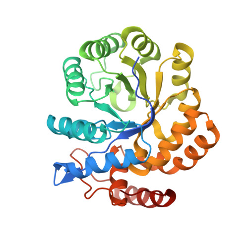 | |
UniProt | |||||
Entity Groups | |||||
| Sequence Clusters | 30% Identity50% Identity70% Identity90% Identity95% Identity100% Identity | ||||
| UniProt Group | Q9PPB4 | ||||
Sequence AnnotationsExpand | |||||
Reference Sequence | |||||
| Ligands 5 Unique | |||||
|---|---|---|---|---|---|
| ID | Chains | Name / Formula / InChI Key | 2D Diagram | 3D Interactions | |
| 3VN (Subject of Investigation/LOI) Download:Ideal Coordinates CCD File | H [auth B], L [auth C] | (2R,5R)-2,5-diamino-2,5-bis(4-aminobutyl)hexanedioic acid C14 H30 N4 O4 XYUHPPVEDOOPDE-ZIAGYGMSSA-N |  | ||
| PGE (Subject of Investigation/LOI) Download:Ideal Coordinates CCD File | G [auth A], J [auth B], Q [auth C], T [auth D] | TRIETHYLENE GLYCOL C6 H14 O4 ZIBGPFATKBEMQZ-UHFFFAOYSA-N |  | ||
| PEG (Subject of Investigation/LOI) Download:Ideal Coordinates CCD File | K [auth B], U [auth D] | DI(HYDROXYETHYL)ETHER C4 H10 O3 MTHSVFCYNBDYFN-UHFFFAOYSA-N |  | ||
| EDO (Subject of Investigation/LOI) Download:Ideal Coordinates CCD File | V [auth D] | 1,2-ETHANEDIOL C2 H6 O2 LYCAIKOWRPUZTN-UHFFFAOYSA-N |  | ||
| MG (Subject of Investigation/LOI) Download:Ideal Coordinates CCD File | E [auth A] F [auth A] I [auth B] M [auth C] N [auth C] | MAGNESIUM ION Mg JLVVSXFLKOJNIY-UHFFFAOYSA-N |  | ||
| Length ( Å ) | Angle ( ˚ ) |
|---|---|
| a = 72.03 | α = 90 |
| b = 84.29 | β = 90 |
| c = 200.94 | γ = 90 |
| Software Name | Purpose |
|---|---|
| XSCALE | data scaling |
| PHENIX | refinement |
| PDB_EXTRACT | data extraction |
| HKL-3000 | data reduction |
| PHENIX | phasing |
| Funding Organization | Location | Grant Number |
|---|---|---|
| Natural Sciences and Engineering Research Council (NSERC, Canada) | Canada | -- |


