7LYJ

SARS-CoV-2 frameshifting pseudoknot RNA


Experimental Data Snapshot

  • Method: X-RAY DIFFRACTION
  • Resolution: 2.11 Å
  • R-Value Free: 0.251 
  • R-Value Work: 0.210 
  • R-Value Observed: 0.214 

wwPDB Validation   3D Report Full Report


This is version 1.2 of the entry. See complete history


Literature

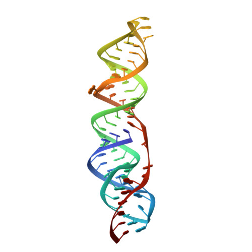
Crystal structure of the severe acute respiratory syndrome coronavirus 2 (SARS-CoV-2) frameshifting pseudoknot.

Jones, C.P.Ferre-D'Amare, A.R.

(2022) RNA 28: 239-249

  • DOI: https://doi.org/10.1261/rna.078825.121
  • Primary Citation of Related Structures:  
    7LYJ, 7MKY

  • PubMed Abstract: 

    SARS-CoV-2 produces two long viral protein precursors from one open reading frame using a highly conserved RNA pseudoknot that enhances programmed -1 ribosomal frameshifting. The 1.3 Å-resolution X-ray structure of the pseudoknot reveals three coaxially stacked helices buttressed by idiosyncratic base triples from loop residues. This structure represents a frameshift-stimulating state that must be deformed by the ribosome and exhibits base-triple-adjacent pockets that could be targeted by future small-molecule therapeutics.


  • Organizational Affiliation

    Biochemistry and Biophysics Center, National Heart, Lung and Blood Institute, Bethesda, Maryland 20892, USA.


Macromolecules
Find similar nucleic acids by:  (by identity cutoff)  |  3D Structure
Entity ID: 1
MoleculeChains LengthOrganismImage
RNA (66-MER)66Severe acute respiratory syndrome coronavirus 2
Sequence Annotations
Expand
  • Reference Sequence
Small Molecules
Ligands 3 Unique
IDChains Name / Formula / InChI Key2D Diagram3D Interactions
IRI
Query on IRI

Download Ideal Coordinates CCD File 
B [auth A],
C [auth A]
IRIDIUM HEXAMMINE ION
H18 Ir N6
CGMAOQLDNKCXGK-UHFFFAOYSA-N
K
Query on K

Download Ideal Coordinates CCD File 
P [auth A],
Q [auth A]
POTASSIUM ION
K
NPYPAHLBTDXSSS-UHFFFAOYSA-N
MG
Query on MG

Download Ideal Coordinates CCD File 
D [auth A]
E [auth A]
F [auth A]
G [auth A]
H [auth A]
D [auth A],
E [auth A],
F [auth A],
G [auth A],
H [auth A],
I [auth A],
J [auth A],
K [auth A],
L [auth A],
M [auth A],
N [auth A],
O [auth A]
MAGNESIUM ION
Mg
JLVVSXFLKOJNIY-UHFFFAOYSA-N
Experimental Data & Validation

Experimental Data

  • Method: X-RAY DIFFRACTION
  • Resolution: 2.11 Å
  • R-Value Free: 0.251 
  • R-Value Work: 0.210 
  • R-Value Observed: 0.214 
  • Space Group: C 1 2 1
Unit Cell:
Length ( Å )Angle ( ˚ )
a = 115.465α = 90
b = 39.477β = 98.53
c = 39.398γ = 90
Software Package:
Software NamePurpose
Aimlessdata scaling
PHENIXrefinement
PDB_EXTRACTdata extraction
XDSdata reduction
AutoSolphasing

Structure Validation

View Full Validation Report


Entry History 

Deposition Data

Revision History  (Full details and data files)

  • Version 1.0: 2021-05-26
    Type: Initial release
  • Version 1.1: 2021-12-08
    Changes: Database references
  • Version 1.2: 2022-02-02
    Changes: Database references