7LXE

ENAH EVH1 domain bound to peptide from ABI1


Experimental Data Snapshot

  • Method: X-RAY DIFFRACTION
  • Resolution: 1.88 Å
  • R-Value Free: 0.276 
  • R-Value Work: 0.245 
  • R-Value Observed: 0.248 

wwPDB Validation   3D Report Full Report


This is version 1.2 of the entry. See complete history


Literature

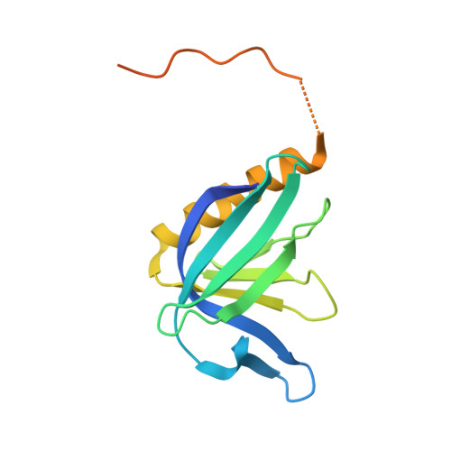
Native proline-rich motifs exploit sequence context to target actin-remodeling Ena/VASP protein ENAH.

Hwang, T.Parker, S.S.Hill, S.M.Grant, R.A.Ilunga, M.W.Sivaraman, V.Mouneimne, G.Keating, A.E.

(2022) Elife 11

  • DOI: https://doi.org/10.7554/eLife.70680
  • Primary Citation of Related Structures:  
    7LXE

  • PubMed Abstract: 

    The human proteome is replete with short linear motifs (SLiMs) of four to six residues that are critical for protein-protein interactions, yet the importance of the sequence surrounding such motifs is underexplored. We devised a proteomic screen to examine the influence of SLiM sequence context on protein-protein interactions. Focusing on the EVH1 domain of human ENAH, an actin regulator that is highly expressed in invasive cancers, we screened 36-residue proteome-derived peptides and discovered new interaction partners of ENAH and diverse mechanisms by which context influences binding. A pocket on the ENAH EVH1 domain that has diverged from other Ena/VASP paralogs recognizes extended SLiMs and favors motif-flanking proline residues. Many high-affinity ENAH binders that contain two proline-rich SLiMs use a noncanonical site on the EVH1 domain for binding and display a thermodynamic signature consistent with the two-motif chain engaging a single domain. We also found that photoreceptor cilium actin regulator (PCARE) uses an extended 23-residue region to obtain a higher affinity than any known ENAH EVH1-binding motif. Our screen provides a way to uncover the effects of proteomic context on motif-mediated binding, revealing diverse mechanisms of control over EVH1 interactions and establishing that SLiMs can't be fully understood outside of their native context.


  • Organizational Affiliation

    Department of Biology, Massachusetts Institute of Technology, Cambridge, United States.


Macromolecules
Find similar proteins by:  (by identity cutoff)  |  3D Structure
Entity ID: 1
MoleculeChains Sequence LengthOrganismDetailsImage
Protein enabled homolog,Abl interactor 1156Homo sapiensMutation(s): 0 
Gene Names: ENAHABI1
UniProt & NIH Common Fund Data Resources
Find proteins for Q8N8S7 (Homo sapiens)
Explore Q8N8S7 
Go to UniProtKB:  Q8N8S7
PHAROS:  Q8N8S7
GTEx:  ENSG00000154380 
Find proteins for Q8IZP0 (Homo sapiens)
Explore Q8IZP0 
Go to UniProtKB:  Q8IZP0
PHAROS:  Q8IZP0
GTEx:  ENSG00000136754 
Entity Groups  
Sequence Clusters30% Identity50% Identity70% Identity90% Identity95% Identity100% Identity
UniProt GroupsQ8IZP0Q8N8S7
Sequence Annotations
Expand
  • Reference Sequence
Experimental Data & Validation

Experimental Data

  • Method: X-RAY DIFFRACTION
  • Resolution: 1.88 Å
  • R-Value Free: 0.276 
  • R-Value Work: 0.245 
  • R-Value Observed: 0.248 
  • Space Group: C 2 2 21
Unit Cell:
Length ( Å )Angle ( ˚ )
a = 56.067α = 90
b = 77.485β = 90
c = 66.167γ = 90
Software Package:
Software NamePurpose
PHENIXrefinement
Aimlessdata scaling
PDB_EXTRACTdata extraction
XDSdata reduction
PHASERphasing

Structure Validation

View Full Validation Report


Entry History & Funding Information

Deposition Data


Funding OrganizationLocationGrant Number
National Institutes of Health/National Institute of General Medical Sciences (NIH/NIGMS)United StatesGM129007

Revision History  (Full details and data files)

  • Version 1.0: 2022-01-19
    Type: Initial release
  • Version 1.1: 2022-08-10
    Changes: Database references
  • Version 1.2: 2023-10-18
    Changes: Data collection, Refinement description