7LUU | pdb_00007luu

Kinetic and Structural Characterization of the First B3 Metallo-beta-Lactamase with an Active Site Glutamic Acid


Experimental Data Snapshot

  • Method: X-RAY DIFFRACTION
  • Resolution: 1.68 Å
  • R-Value Free: 
    0.170 (Depositor), 0.158 (DCC) 
  • R-Value Work: 
    0.141 (Depositor) 
  • R-Value Observed: 
    0.143 (Depositor) 

Starting Model: experimental
View more details

wwPDB Validation 3D Report Full Report

Validation slider image for 7LUU

This is version 1.3 of the entry. See complete history

Literature

Kinetic and Structural Characterization of the First B3 Metallo-beta-Lactamase with an Active-Site Glutamic Acid.

Wilson, L.A.Knaven, E.G.Morris, M.T.Monteiro Pedroso, M.Schofield, C.J.Bruck, T.B.Boden, M.Waite, D.W.Hugenholtz, P.Guddat, L.Schenk, G.

(2021) Antimicrob Agents Chemother 65: e0093621-e0093621

  • DOI: https://doi.org/10.1128/AAC.00936-21
  • Primary Citation Related Structures: 
    7LUU

  • PubMed Abstract: 

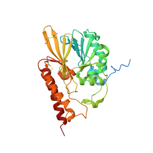
    The structural diversity in metallo-β-lactamases (MBLs), especially in the vicinity of the active site, has been a major hurdle in the development of clinically effective inhibitors. Representatives from three variants of the B3 MBL subclass, containing either the canonical HHH/DHH active-site motif (present in the majority of MBLs in this subclass) or the QHH/DHH (B3-Q) or HRH/DQK (B3-RQK) variations, were reported previously. Here, we describe the structure and kinetic properties of the first example (SIE-1) of a fourth variant containing the EHH/DHH active-site motif (B3-E). SIE-1 was identified in the hexachlorocyclohexane-degrading bacterium Sphingobium indicum, and kinetic analyses demonstrate that although it is active against a wide range of antibiotics, its efficiency is lower than that of other B3 MBLs but has increased efficiency toward cephalosporins relative to other β-lactam substrates. The overall fold of SIE-1 is characteristic of the MBLs; the notable variation is observed in the Zn1 site due to the replacement of the canonical His116 by a glutamate. The unusual preference of SIE-1 for cephalosporins and its occurrence in a widespread environmental organism suggest the scope for increased MBL-mediated β-lactam resistance. Thus, it is relevant to include SIE-1 in MBL inhibitor design studies to widen the therapeutic scope of much needed antiresistance drugs.


  • Organizational Affiliation
    • School of Chemistry and Molecular Biosciences, The University of Queenslandgrid.1003.2, St. Lucia, QLD, Brisbane, Australia.

Macromolecule Content 

  • Total Structure Weight: 34.8 kDa 
  • Atom Count: 2,472 
  • Modeled Residue Count: 266 
  • Deposited Residue Count: 307 
  • Unique protein chains: 1

Macromolecules

Find similar proteins by:|  3D Structure
Entity ID: 1
MoleculeChains  Sequence LengthOrganismDetailsImage
Subclass B3 metallo-beta-lactamase307Sphingobium indicum B90AMutation(s): 0 
Gene Names: SIDU_11385
UniProt
Find proteins for A0A1L5BQA7 (Sphingobium indicum (strain DSM 16412 / CCM 7286 / MTCC 6364 / B90A))
Explore A0A1L5BQA7 
Go to UniProtKB:  A0A1L5BQA7
Entity Groups
Sequence Clusters30% Identity50% Identity70% Identity90% Identity95% Identity100% Identity
UniProt GroupA0A1L5BQA7
Sequence Annotations
Expand
Reference Sequence

Small Molecules

Ligands 3 Unique
IDChains Name / Formula / InChI Key2D Diagram3D Interactions
SO4

Query on SO4


Download:Ideal Coordinates CCD File
AA [auth A],
D [auth A],
E [auth A]
SULFATE ION
O4 S
QAOWNCQODCNURD-UHFFFAOYSA-L
GOL

Query on GOL


Download:Ideal Coordinates CCD File
F [auth A]
G [auth A]
H [auth A]
I [auth A]
J [auth A]
F [auth A],
G [auth A],
H [auth A],
I [auth A],
J [auth A],
K [auth A],
L [auth A],
M [auth A],
N [auth A],
O [auth A],
P [auth A],
Q [auth A],
R [auth A],
S [auth A],
T [auth A],
U [auth A],
V [auth A],
W [auth A],
X [auth A],
Y [auth A],
Z [auth A]
GLYCEROL
C3 H8 O3
PEDCQBHIVMGVHV-UHFFFAOYSA-N
ZN
(Subject of Investigation/LOI)

Query on ZN


Download:Ideal Coordinates CCD File
B [auth A],
C [auth A]
ZINC ION
Zn
PTFCDOFLOPIGGS-UHFFFAOYSA-N

Experimental Data & Validation

Experimental Data

  • Method: X-RAY DIFFRACTION
  • Resolution: 1.68 Å
  • R-Value Free:  0.170 (Depositor), 0.158 (DCC) 
  • R-Value Work:  0.141 (Depositor) 
  • R-Value Observed: 0.143 (Depositor) 
Space Group: P 21 21 2
Unit Cell:
Length ( Å )Angle ( ˚ )
a = 62.64α = 90
b = 75.42β = 90
c = 56.67γ = 90
Software Package:
Software NamePurpose
Aimlessdata scaling
PHENIXrefinement
PDB_EXTRACTdata extraction
XDSdata reduction
PHASERphasing

Structure Validation

View Full Validation Report


Entry History 

& Funding Information

Deposition Data


Funding OrganizationLocationGrant Number
National Health and Medical Research Council (NHMRC, Australia)AustraliaAPP1084778

Revision History  (Full details and data files)

  • Version 1.0: 2021-07-28
    Type: Initial release
  • Version 1.1: 2022-02-09
    Changes: Database references
  • Version 1.2: 2023-10-18
    Changes: Data collection, Refinement description
  • Version 1.3: 2024-11-06
    Changes: Structure summary