7LRF | pdb_00007lrf

Netrin-1 in complex with SOS


Experimental Data Snapshot

  • Method: X-RAY DIFFRACTION
  • Resolution: 3.21 Å
  • R-Value Free: 
    0.281 (Depositor), 0.279 (DCC) 
  • R-Value Work: 
    0.226 (Depositor), 0.226 (DCC) 
  • R-Value Observed: 
    0.229 (Depositor) 

Starting Model: experimental
View more details

wwPDB Validation 3D Report Full Report

Validation slider image for 7LRF

This is version 1.3 of the entry. See complete history

Literature

The dynamic nature of netrin-1 and the structural basis for glycosaminoglycan fragment-induced filament formation.

Meier, M.Gupta, M.Akgul, S.McDougall, M.Imhof, T.Nikodemus, D.Reuten, R.Moya-Torres, A.To, V.Ferens, F.Heide, F.Padilla-Meier, G.P.Kukura, P.Huang, W.Gerisch, B.Morgelin, M.Poole, K.Antebi, A.Koch, M.Stetefeld, J.

(2023) Nat Commun 14: 1226-1226

  • DOI: https://doi.org/10.1038/s41467-023-36692-w
  • Primary Citation Related Structures: 
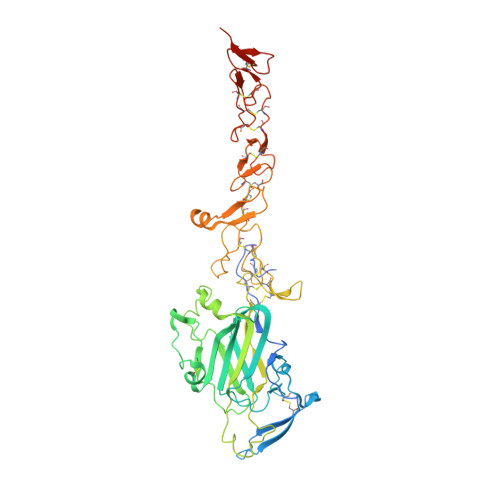
    7LER, 7LRF

  • PubMed Abstract: 

    Netrin-1 is a bifunctional chemotropic guidance cue that plays key roles in diverse cellular processes including axon pathfinding, cell migration, adhesion, differentiation, and survival. Here, we present a molecular understanding of netrin-1 mediated interactions with glycosaminoglycan chains of diverse heparan sulfate proteoglycans (HSPGs) and short heparin oligosaccharides. Whereas interactions with HSPGs act as platform to co-localise netrin-1 close to the cell surface, heparin oligosaccharides have a significant impact on the highly dynamic behaviour of netrin-1. Remarkably, the monomer-dimer equilibrium of netrin-1 in solution is abolished in the presence of heparin oligosaccharides and replaced with highly hierarchical and distinct super assemblies leading to unique, yet unknown netrin-1 filament formation. In our integrated approach we provide a molecular mechanism for the filament assembly which opens fresh paths towards a molecular understanding of netrin-1 functions.


  • Organizational Affiliation
    • Department of Chemistry, University of Manitoba, Winnipeg, Canada.

Macromolecule Content 

  • Total Structure Weight: 105.49 kDa 
  • Atom Count: 6,726 
  • Modeled Residue Count: 829 
  • Deposited Residue Count: 886 
  • Unique protein chains: 1

Macromolecules

Find similar proteins by:|  3D Structure
Entity ID: 1
MoleculeChains  Sequence LengthOrganismDetailsImage
Netrin-1
A, B
443Gallus gallusMutation(s): 0 
Gene Names: NTN1
UniProt
Find proteins for Q90922 (Gallus gallus)
Explore Q90922 
Go to UniProtKB:  Q90922
Entity Groups
Sequence Clusters30% Identity50% Identity70% Identity90% Identity95% Identity100% Identity
UniProt GroupQ90922
Glycosylation
Glycosylation Sites: 4
Sequence Annotations
Expand
Reference Sequence

Oligosaccharides

Help  
Entity ID: 2
MoleculeChains Length2D Diagram GlycosylationD Interactions
beta-D-mannopyranose-(1-4)-2-acetamido-2-deoxy-beta-D-glucopyranose-(1-4)-2-acetamido-2-deoxy-beta-D-glucopyranose
C
3N-Glycosylation
Glycosylation Resources
GlyTouCan: G15407YE
GlyCosmos: G15407YE
GlyGen: G15407YE
Entity ID: 3
MoleculeChains Length2D Diagram GlycosylationD Interactions
2-acetamido-2-deoxy-beta-D-glucopyranose-(1-4)-2-acetamido-2-deoxy-beta-D-glucopyranose
D, E, F, G, H
D, E, F, G, H, I, J
2N-Glycosylation
Glycosylation Resources
GlyTouCan: G42666HT
GlyCosmos: G42666HT
GlyGen: G42666HT
Entity ID: 4
MoleculeChains Length2D Diagram GlycosylationD Interactions
1,3,4,6-tetra-O-sulfo-beta-D-fructofuranose-(2-1)-2,3,4,6-tetra-O-sulfonato-alpha-D-glucopyranose
K, L
2N/A
Glycosylation Resources
GlyTouCan: G97555GV
GlyCosmos: G97555GV

Small Molecules

Ligands 6 Unique
IDChains Name / Formula / InChI Key2D Diagram3D Interactions
NHE

Query on NHE


Download:Ideal Coordinates CCD File
T [auth B]2-[N-CYCLOHEXYLAMINO]ETHANE SULFONIC ACID
C8 H17 N O3 S
MKWKNSIESPFAQN-UHFFFAOYSA-N
PEG

Query on PEG


Download:Ideal Coordinates CCD File
P [auth A],
U [auth B]
DI(HYDROXYETHYL)ETHER
C4 H10 O3
MTHSVFCYNBDYFN-UHFFFAOYSA-N
EDO

Query on EDO


Download:Ideal Coordinates CCD File
M [auth A],
N [auth A],
O [auth A],
S [auth B]
1,2-ETHANEDIOL
C2 H6 O2
LYCAIKOWRPUZTN-UHFFFAOYSA-N
CA

Query on CA


Download:Ideal Coordinates CCD File
Q [auth A],
V [auth B]
CALCIUM ION
Ca
BHPQYMZQTOCNFJ-UHFFFAOYSA-N
CL

Query on CL


Download:Ideal Coordinates CCD File
W [auth B]CHLORIDE ION
Cl
VEXZGXHMUGYJMC-UHFFFAOYSA-M
NA

Query on NA


Download:Ideal Coordinates CCD File
R [auth A]SODIUM ION
Na
FKNQFGJONOIPTF-UHFFFAOYSA-N

Biologically Interesting Molecules (External Reference) 

1 Unique

Experimental Data & Validation

Experimental Data

  • Method: X-RAY DIFFRACTION
  • Resolution: 3.21 Å
  • R-Value Free:  0.281 (Depositor), 0.279 (DCC) 
  • R-Value Work:  0.226 (Depositor), 0.226 (DCC) 
  • R-Value Observed: 0.229 (Depositor) 
Space Group: P 21 21 21
Unit Cell:
Length ( Å )Angle ( ˚ )
a = 75.105α = 90
b = 80.152β = 90
c = 241.702γ = 90
Software Package:
Software NamePurpose
PHENIXrefinement
Aimlessdata scaling
PDB_EXTRACTdata extraction
XDSdata reduction
PHASERphasing

Structure Validation

View Full Validation Report


Entry History 

& Funding Information

Deposition Data


Funding OrganizationLocationGrant Number
Canadian Institutes of Health Research (CIHR)CanadaRPA-109759

Revision History  (Full details and data files)

  • Version 1.0: 2022-02-23
    Type: Initial release
  • Version 1.1: 2023-03-15
    Changes: Database references
  • Version 1.2: 2023-10-25
    Changes: Data collection, Refinement description
  • Version 1.3: 2024-10-16
    Changes: Structure summary