Crystal Structure of Escherichia coli Agmatinase: Catalytic Mechanism and Residues Relevant for Substrate Specificity.
Maturana, P., Orellana, M.S., Herrera, S.M., Martinez, I., Figueroa, M., Martinez-Oyanedel, J., Castro-Fernandez, V., Uribe, E.(2021) Int J Mol Sci 22
- PubMed: 33946272 Search on PubMedSearch on PubMed Central
- DOI: https://doi.org/10.3390/ijms22094769
- Primary Citation Related Structures:
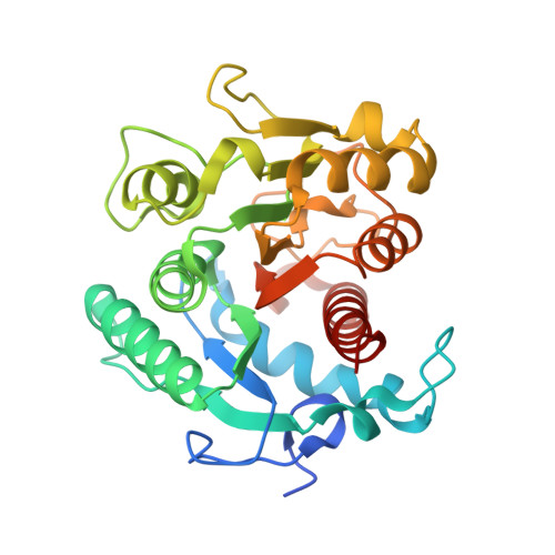
7LOL, 7LOX - PubMed Abstract:
Agmatine is the product of the decarboxylation of L-arginine by the enzyme arginine decarboxylase. This amine has been attributed to neurotransmitter functions, anticonvulsant, anti-neurotoxic, and antidepressant in mammals and is a potential therapeutic agent for diseases such as Alzheimer's, Parkinson's, and cancer. Agmatinase enzyme hydrolyze agmatine into urea and putrescine, which belong to one of the pathways producing polyamines, essential for cell proliferation. Agmatinase from Escherichia coli (EcAGM) has been widely studied and kinetically characterized, described as highly specific for agmatine. In this study, we analyze the amino acids involved in the high specificity of EcAGM, performing a series of mutations in two loops critical to the active-site entrance. Two structures in different space groups were solved by X-ray crystallography, one at low resolution (3.2 Å), including a guanidine group; and other at high resolution (1.8 Å) which presents urea and agmatine in the active site. These structures made it possible to understand the interface interactions between subunits that allow the hexameric state and postulate a catalytic mechanism according to the Mn 2+ and urea/guanidine binding site. Molecular dynamics simulations evaluated the conformational dynamics of EcAGM and residues participating in non-binding interactions. Simulations showed the high dynamics of loops of the active site entrance and evidenced the relevance of Trp68, located in the adjacent subunit, to stabilize the amino group of agmatine by cation-pi interaction. These results allow to have a structural view of the best-kinetic characterized agmatinase in literature up to now.
- Departamento de Biología, Facultad de Ciencias, Universidad de Chile, Ñuñoa 7800003, Santiago, Chile.
Organizational Affiliation: