7LM9 | pdb_00007lm9

Crystal structure of SARS-CoV spike protein receptor-binding domain in complex with a cross-neutralizing antibody CV38-142 Fab isolated from COVID-19 patient


Experimental Data Snapshot

  • Method: X-RAY DIFFRACTION
  • Resolution: 1.53 Å
  • R-Value Free: 
    0.192 (Depositor), 0.193 (DCC) 
  • R-Value Work: 
    0.170 (Depositor), 0.171 (DCC) 
  • R-Value Observed: 
    0.171 (Depositor) 

Starting Models: experimental
View more details

wwPDB Validation 3D Report Full Report

Validation slider image for 7LM9

This is version 1.3 of the entry. See complete history. 

Literature

A combination of cross-neutralizing antibodies synergizes to prevent SARS-CoV-2 and SARS-CoV pseudovirus infection.

Liu, H., Yuan, M., Huang, D., Bangaru, S., Zhao, F., Lee, C.D., Peng, L., Barman, S., Zhu, X., Nemazee, D., Burton, D.R., van Gils, M.J., Sanders, R.W., Kornau, H.C., Reincke, S.M., Pruss, H., Kreye, J., Wu, N.C., Ward, A.B., Wilson, I.A.

(2021) Cell Host Microbe 29: 806-818.e6

  • DOI: https://doi.org/10.1016/j.chom.2021.04.005
  • Primary Citation Related Structures: 
    7LM8, 7LM9

  • PubMed Abstract: 

    Coronaviruses have caused several human epidemics and pandemics including the ongoing coronavirus disease 2019 (COVID-19). Prophylactic vaccines and therapeutic antibodies have already shown striking effectiveness against COVID-19. Nevertheless, concerns remain about antigenic drift in SARS-CoV-2 as well as threats from other sarbecoviruses. Cross-neutralizing antibodies to SARS-related viruses provide opportunities to address such concerns. Here, we report on crystal structures of a cross-neutralizing antibody, CV38-142, in complex with the receptor-binding domains from SARS-CoV-2 and SARS-CoV. Recognition of the N343 glycosylation site and water-mediated interactions facilitate cross-reactivity of CV38-142 to SARS-related viruses, allowing the antibody to accommodate antigenic variation in these viruses. CV38-142 synergizes with other cross-neutralizing antibodies, notably COVA1-16, to enhance neutralization of SARS-CoV and SARS-CoV-2, including circulating variants of concern B.1.1.7 and B.1.351. Overall, this study provides valuable information for vaccine and therapeutic design to address current and future antigenic drift in SARS-CoV-2 and to protect against zoonotic SARS-related coronaviruses.


  • Organizational Affiliation: 
    • Department of Integrative Structural and Computational Biology, The Scripps Research Institute, La Jolla, CA 92037, USA.

Macromolecule Content 

  • Total Structure Weight: 75.15 kDa 
  • Atom Count: 5,619 
  • Modeled Residue Count: 634 
  • Deposited Residue Count: 673 
  • Unique protein chains: 3

Macromolecules

Find similar proteins by:|  3D Structure
Entity ID: 1
MoleculeChains  Sequence LengthOrganismDetailsImage
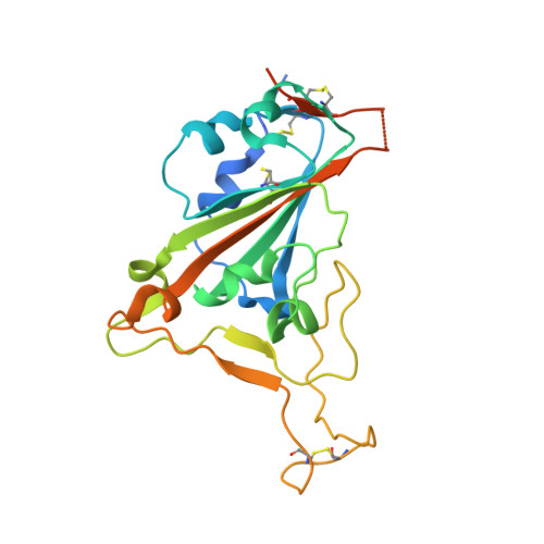
Spike glycoprotein230SARS coronavirus MA15Mutation(s): 0 
UniProt
Find proteins for D2E265 (SARS coronavirus MA15)
Explore D2E265 
Go to UniProtKB:  D2E265
Entity Groups
Sequence Clusters30% Identity50% Identity70% Identity90% Identity95% Identity100% Identity
UniProt GroupD2E265
Glycosylation
Glycosylation Sites: 1
Sequence Annotations
Expand
Reference Sequence
Find similar proteins by:|  3D Structure
Entity ID: 2
MoleculeChains  Sequence LengthOrganismDetailsImage
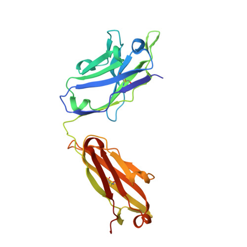
CV38-142 Fab heavy chainB [auth H]226Homo sapiensMutation(s): 0 
Entity Groups
Sequence Clusters30% Identity50% Identity70% Identity90% Identity95% Identity100% Identity
Sequence Annotations
Expand
Reference Sequence
Find similar proteins by:|  3D Structure
Entity ID: 3
MoleculeChains  Sequence LengthOrganismDetailsImage
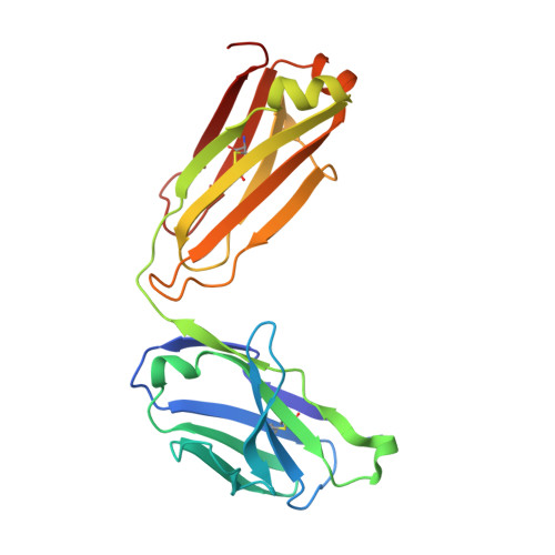
CV38-142 Fab light chainC [auth L]217Homo sapiensMutation(s): 0 
Entity Groups
Sequence Clusters30% Identity50% Identity70% Identity90% Identity95% Identity100% Identity
Sequence Annotations
Expand
Reference Sequence

Oligosaccharides

Help  
Entity ID: 4
MoleculeChains Length2D Diagram GlycosylationD Interactions
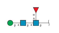
beta-D-mannopyranose-(1-4)-2-acetamido-2-deoxy-beta-D-glucopyranose-(1-4)-[alpha-L-fucopyranose-(1-6)]2-acetamido-2-deoxy-beta-D-glucopyranoseD [auth B]4N-Glycosylation
Glycosylation Resources
GlyTouCan: G32152BH
GlyCosmos: G32152BH
GlyGen: G32152BH

Small Molecules

Ligands 1 Unique
IDChains Name / Formula / InChI Key2D Diagram3D Interactions
EDO

Query on EDO


Download:Ideal Coordinates CCD File
E [auth A]
F [auth A]
G [auth A]
H [auth A]
I [auth H]
E [auth A],
F [auth A],
G [auth A],
H [auth A],
I [auth H],
J [auth H],
K [auth L],
L,
M [auth L],
N [auth L]
1,2-ETHANEDIOL
C2 H6 O2
LYCAIKOWRPUZTN-UHFFFAOYSA-N

Experimental Data & Validation

Experimental Data

  • Method: X-RAY DIFFRACTION
  • Resolution: 1.53 Å
  • R-Value Free:  0.192 (Depositor), 0.193 (DCC) 
  • R-Value Work:  0.170 (Depositor), 0.171 (DCC) 
  • R-Value Observed: 0.171 (Depositor) 
Space Group: C 1 2 1
Unit Cell:
Length ( Å )Angle ( ˚ )
a = 237.997α = 90
b = 71.877β = 90.78
c = 49.241γ = 90
Software Package:
Software NamePurpose
PHENIXrefinement
HKL-2000data scaling
PDB_EXTRACTdata extraction
HKL-2000data reduction
PHASERphasing

Structure Validation

View Full Validation Report


Entry History 

& Funding Information

Deposition Data


Funding OrganizationLocationGrant Number
Bill & Melinda Gates FoundationUnited StatesOPP1170236
Bill & Melinda Gates FoundationUnited StatesINV-004923

Revision History  (Full details and data files)

  • Version 1.0: 2021-03-31
    Type: Initial release
  • Version 1.1: 2022-02-09
    Changes: Database references
  • Version 1.2: 2023-10-18
    Changes: Data collection, Database references, Refinement description
  • Version 1.3: 2024-11-20
    Changes: Structure summary