7LL2 | pdb_00007ll2

Cryo-EM structure of BG505 DS-SOSIP in complex with Glycan276-Dependent Broadly Neutralizing Antibody VRC33.01 Fab


Experimental Data Snapshot

  • Method: ELECTRON MICROSCOPY
  • Resolution: 3.73 Å
  • Aggregation State: PARTICLE 
  • Reconstruction Method: SINGLE PARTICLE 

Starting Models: experimental
View more details

wwPDB Validation 3D Report Full Report

Validation slider image for 7LL2

This is version 1.2 of the entry. See complete history. 

Literature

Structural basis of glycan276-dependent recognition by HIV-1 broadly neutralizing antibodies.

Cottrell, C.A., Manne, K., Kong, R., Wang, S., Zhou, T., Chuang, G.Y., Edwards, R.J., Henderson, R., Janowska, K., Kopp, M., Lin, B.C., Louder, M.K., Olia, A.S., Rawi, R., Shen, C.H., Taft, J.D., Torres, J.L., Wu, N.R., Zhang, B., Doria-Rose, N.A., Cohen, M.S., Haynes, B.F., Shapiro, L., Ward, A.B., Acharya, P., Mascola, J.R., Kwong, P.D.

(2021) Cell Rep 37: 109922-109922

  • DOI: https://doi.org/10.1016/j.celrep.2021.109922
  • Primary Citation Related Structures: 
    7LG6, 7LL1, 7LL2, 7LLK

  • PubMed Abstract: 

    Recognition of N-linked glycan at residue N276 (glycan276) at the periphery of the CD4-binding site (CD4bs) on the HIV-envelope trimer is a formidable challenge for many CD4bs-directed antibodies. To understand how this glycan can be recognized, here we isolate two lineages of glycan276-dependent CD4bs antibodies. Antibody CH540-VRC40.01 (named for donor-lineage.clone) neutralizes 81% of a panel of 208 diverse strains, while antibody CH314-VRC33.01 neutralizes 45%. Cryo-electron microscopy (cryo-EM) structures of these two antibodies and 179NC75, a previously identified glycan276-dependent CD4bs antibody, in complex with HIV-envelope trimer reveal substantially different modes of glycan276 recognition. Despite these differences, binding of glycan276-dependent antibodies maintains a glycan276 conformation similar to that observed in the absence of glycan276-binding antibodies. By contrast, glycan276-independent CD4bs antibodies, such as VRC01, displace glycan276 upon binding. These results provide a foundation for understanding antibody recognition of glycan276 and suggest its presence may be crucial for priming immunogens seeking to initiate broad CD4bs recognition.


  • Organizational Affiliation: 
    • IAVI Neutralizing Antibody Center, Consortium for HIV/AIDS Vaccine Development (CHAVD), Department of Integrative Structural and Computational Biology, The Scripps Research Institute, La Jolla, CA 92037, USA.

Macromolecule Content 

  • Total Structure Weight: 379.89 kDa 
  • Atom Count: 25,512 
  • Modeled Residue Count: 3,060 
  • Deposited Residue Count: 3,204 
  • Unique protein chains: 4

Macromolecules

Find similar proteins by:|  3D Structure
Entity ID: 1
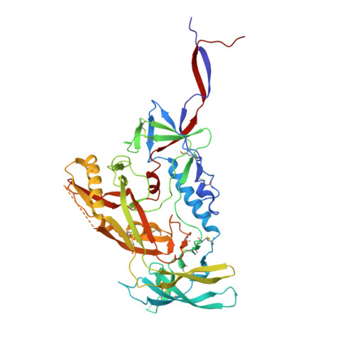
MoleculeChains  Sequence LengthOrganismDetailsImage
Envelope glycoprotein gp120A,
E [auth C],
I [auth E]
473Human immunodeficiency virus 1Mutation(s): 0 
Gene Names: env
UniProt
Find proteins for Q2N0S6 (Human immunodeficiency virus type 1)
Explore Q2N0S6 
Go to UniProtKB:  Q2N0S6
Entity Groups
Sequence Clusters30% Identity50% Identity70% Identity90% Identity95% Identity100% Identity
UniProt GroupQ2N0S6
Glycosylation
Glycosylation Sites: 15
Sequence Annotations
Expand
Reference Sequence
Find similar proteins by:|  3D Structure
Entity ID: 2
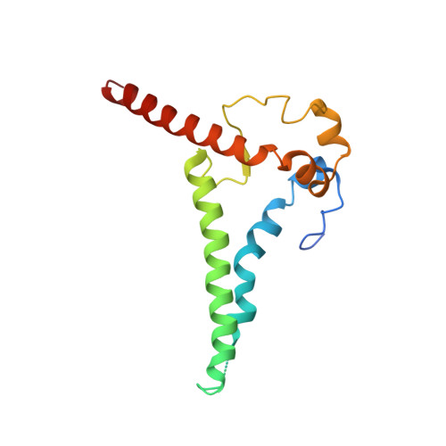
MoleculeChains  Sequence LengthOrganismDetailsImage
Envelope glycoprotein gp41B,
F [auth D],
J [auth F]
153Human immunodeficiency virus 1Mutation(s): 0 
Gene Names: env
UniProt
Find proteins for Q2N0S7 (Human immunodeficiency virus type 1)
Explore Q2N0S7 
Go to UniProtKB:  Q2N0S7
Entity Groups
Sequence Clusters30% Identity50% Identity70% Identity90% Identity95% Identity100% Identity
UniProt GroupQ2N0S7
Sequence Annotations
Expand
Reference Sequence
Find similar proteins by:|  3D Structure
Entity ID: 3
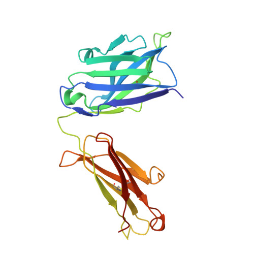
MoleculeChains  Sequence LengthOrganismDetailsImage
VRC33.01 Fab Heavy chainC [auth H],
G [auth I],
K
225Homo sapiensMutation(s): 0 
Entity Groups
Sequence Clusters30% Identity50% Identity70% Identity90% Identity95% Identity100% Identity
Sequence Annotations
Expand
Reference Sequence
Find similar proteins by:|  3D Structure
Entity ID: 4
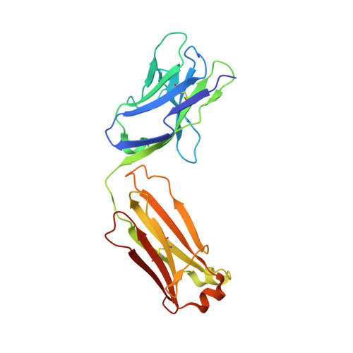
MoleculeChains  Sequence LengthOrganismDetailsImage
VRC33.01 Fab Light chainD [auth L],
H [auth J],
L [auth M]
217Homo sapiensMutation(s): 0 
Entity Groups
Sequence Clusters30% Identity50% Identity70% Identity90% Identity95% Identity100% Identity
Sequence Annotations
Expand
Reference Sequence

Oligosaccharides

Help  
Entity ID: 6
MoleculeChains Length2D Diagram GlycosylationD Interactions
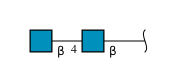
2-acetamido-2-deoxy-beta-D-glucopyranose-(1-4)-2-acetamido-2-deoxy-beta-D-glucopyranoseJA [auth j],
N,
Y
2N-Glycosylation
Glycosylation Resources
GlyTouCan: G42666HT
GlyCosmos: G42666HT
GlyGen: G42666HT
Entity ID: 7
MoleculeChains Length2D Diagram GlycosylationD Interactions
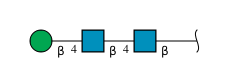
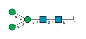
alpha-D-mannopyranose-(1-3)-[alpha-D-mannopyranose-(1-6)]beta-D-mannopyranose-(1-4)-2-acetamido-2-deoxy-beta-D-glucopyranose-(1-4)-2-acetamido-2-deoxy-beta-D-glucopyranose
DA [auth d],
FA [auth f],
OA [auth o],
QA [auth q],
S,
DA [auth d],
FA [auth f],
OA [auth o],
QA [auth q],
S,
U
5N-Glycosylation
Glycosylation Resources
GlyTouCan: G22768VO
GlyCosmos: G22768VO
GlyGen: G22768VO
Entity ID: 8
MoleculeChains Length2D Diagram GlycosylationD Interactions
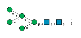
alpha-D-mannopyranose-(1-3)-[alpha-D-mannopyranose-(1-6)]alpha-D-mannopyranose-(1-6)-[alpha-D-mannopyranose-(1-3)]beta-D-mannopyranose-(1-4)-2-acetamido-2-deoxy-beta-D-glucopyranose-(1-4)-2-acetamido-2-deoxy-beta-D-glucopyranoseEA [auth e],
PA [auth p],
T
7N-Glycosylation
Glycosylation Resources
GlyTouCan: G55220VL
GlyCosmos: G55220VL
GlyGen: G55220VL

Small Molecules

Ligands 1 Unique
IDChains Name / Formula / InChI Key2D Diagram3D Interactions
NAG

Query on NAG


Download:Ideal Coordinates CCD File
AB [auth C]
BB [auth E]
CB [auth E]
DB [auth E]
EB [auth E]
AB [auth C],
BB [auth E],
CB [auth E],
DB [auth E],
EB [auth E],
TA [auth A],
UA [auth A],
VA [auth A],
WA [auth A],
XA [auth C],
YA [auth C],
ZA [auth C]
2-acetamido-2-deoxy-beta-D-glucopyranose
C8 H15 N O6
OVRNDRQMDRJTHS-FMDGEEDCSA-N

Experimental Data & Validation

Experimental Data

  • Method: ELECTRON MICROSCOPY
  • Resolution: 3.73 Å
  • Aggregation State: PARTICLE 
  • Reconstruction Method: SINGLE PARTICLE 
EM Software:
TaskSoftware PackageVersion
RECONSTRUCTIONcryoSPARC
MODEL REFINEMENTCoot
MODEL REFINEMENTPHENIX
MODEL REFINEMENTISOLDE

Structure Validation

View Full Validation Report


Entry History 

& Funding Information

Deposition Data


Funding OrganizationLocationGrant Number
National Institutes of Health/National Institute Of Allergy and Infectious Diseases (NIH/NIAID)United StatesAI45687

Revision History  (Full details and data files)

  • Version 1.0: 2021-03-17
    Type: Initial release
  • Version 1.1: 2021-11-17
    Changes: Database references
  • Version 1.2: 2024-10-30
    Changes: Data collection, Refinement description, Structure summary